| Name: | Rabbit polyclonal antibody to ubiquitin |
| Immunogen: | Purified ubiquitin conjugated with glutaraldehyde to KLH |
| HGNC Name: | UBB, UBC, UBA52, RPS27A |
| UniProt: | P0CG48, P0CG47, P62987, P62979 |
| Molecular Weight: | 8.5kDa |
| Host: | Rabbit |
| Isotype: | |
| Species Cross-Reactivity: | Human, rat, mouse |
| RRID: | AB_2253901 |
| Format: | Supplied as an aliquot of serum plus 5mM NaN3 |
| Applications: | WB, ICC/IF, IHC |
| Recommended Dilutions: | WB: 1:5,000-1:10,000. IF/ICC and IHC: 1:500-1:1,000. |
| Storage: | Storage for short term at 4°C recommended, for longer term at -20°C, minimize freeze/thaw cycles |
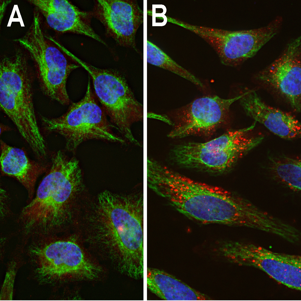
Immunofluorescent analysis of HeLa cells stained with rabbit pAb to ubiquitin, RPCA-Ubi, dilution 1:1,000 in red, and costained with chicken pAb to vimentin, CPCA-Vim, dilution 1:10,000, in green. The blue is DAPI staining of nuclear DNA. [A] Control HeLa cells maintained in normal medium, [B] HeLa cells treated with 10µM of the proteasome inhibitor lactacystin (Lc) for 24 hours. Proteasomal inhibition leads to formation of strongly ubiquitin positive cytoplasmic inclusions. Note the diffuse cytoplasmic ubiquitin staining in control cells and well defined ubiquitin positive inclusions in the Lc treated cells.
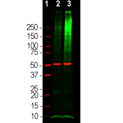
Western blot analysis of HEK293 cell lysates using rabbit pAb to ubiquitin, RPCA-Ubi, dilution 1:5,000 in green and mouse mAb to β-Tubulin, MCA-1B12, dilution 1:10,000, in red, used as a loading control. [1] protein standard (red), [2] cells maintained in normal medium, [3] cells treated with proteasome inhibitor lactacystin (Lc) at 10µM for 16 hours. the lysate was subjected to electrophoresis on a 4-20% SDS-PAGE gel, then electrophoretically transferred to PVDF membranes. The smear detected above the 200kDa standard represents accumulations of ubiquitinated proteins in the Lc treated cells. The prominent band at ~8kDa corresponds to monoubiquitin.
Rabbit Polyclonal Antibody to Ubiquitin
Cat# RPCA-Ubi
Price range: $120.00 through $800.00
Ubiquitin is a globular 76 amino acid protein of about 8.5kDa molecular weight which was discovered by biochemical isolation from bovine thymus tissues (1). The protein was found to be highly conserved in amino acid sequence and was detectable in apparently every cell and tissue type, and, being apparently ubiquitously expressed, became known as ubiquitin. Subsequent work showed that ubiquitin has an important role in the targeting of proteins for proteolytic degradation, but has other important functions (2). Proteins to be degraded are covalently coupled to the C-terminus of ubiquitin as a result of the sequential activity of three families of enzymes, the ubiquitin-activating enzymes (E1s), ubiquitin-conjugating enzymes (E2s), and ubiquitin ligases (E3s). While humans express only two E1 enzymes, there are 35 E2 enzymes and hundreds of E3s. The ubiquitinated protein complex may then be degraded in the proteasome. Ubiquitin becomes covalently bonded to many types of pathological inclusions such as the neurofibrillary tangles of Alzheimer’s disease (3), the Lewy bodies of Parkinson’s disease (4), the Pick bodies of Pick’s disease (5) and many others. Ubiquitin can normally be removed from proteins to which it is bound and reused due to the activity of a large family of deubiquitinating enzymes.
This antibody, RPCA-Ubi, was made against purified bovine blood derived ubiquitin coupled to keyhole limpet hemocyanin with glutaraldehyde (6). The antibody is relatively insensitive to formalin fixation and so can be used on paraffin embedded fixed histological sections of human brain for studies of Alzheimer’s and other neurodegenerative diseases. We also supply a widely used mouse monoclonal antibody to ubiquitin, MCA-Ubi-1. This antibody binds a peptide which is totally conserved in animals, plants and fungi so MCA-Ubi-1 is applicable to studies of a wide variety of species form human to yeast. MCA-Ubi-1 also works on western blots and can be used to study ubiquitinated proteins which typically accumulate as a high molecular weight smear on western blots if the proteastome is inhibited. Mouse select image at left for larger view.

Chromogenic immunostaining of a NBF fixed paraffin embedded human hippocampus section from an Alzheimer’s Disease case. Rabbit pAb to ubiquitin, MCA-Ubi, dilution 1:2,000, detected with DAB (brown) using the Vector Labs ImmPRESS method and reagents with citra buffer retrieval. Hematoxylin (blue) was used as the counterstain. The RPCA-Ubi antibody strongly labels the cytoplasm of diseased neurons as identified by their morphology. Mouse select image for larger view.
1. Goldstein G, et al. Isolation of a polypeptide that has lymphocyte-differentiating properties and is probably represented universally in living cells.
PNAS 72:11-5 (1975).
2. Wilkinson K. The discovery of ubiquitin-dependent proteolysis. PNAS 102:15280-2 (2005).
3. Perry G. et al. Ubiquitin is detected in neurofibrillary tangles and senile plaque neurites of Alzheimer disease brains. PNAS 84:3033-6 (1987).
4. Kuzuhara S, et al. Lewy bodies are ubiquitinated. A light and electron microscopic immunocytochemical study. Acta Neuropathol. 75:345-53 (1988).
5. Murayama S. et al. Immunocytochemical and ultrastructural studies of Pick’s disease. Ann. Neurol. 27:394-405 (1990).
6. Shaw G. Chau V. Ubiquitin and microtubule-associated protein tau immunoreactivity each define distinct structures with differing distributions and solubility properties in Alzheimer brain. PNAS 85:2854-8 (1988).


