| Name: | Mouse monoclonal antibody to Ubiquitin |
| Immunogen: | Purified bovine erythrocyte ubiquitin coupled to KLH with glutaraldehyde |
| HGNC Name: | UBB, UBC, UBA52, RPS27A |
| UniProt: | P0CG48, P0CG47, P62987, P62979 |
| Molecular Weight: | 8.5kDa |
| Host: | Mouse |
| Isotype: | IgG1 heavy, κ light |
| Species Cross-Reactivity: | Human, monkey, rat, mouse, horse, cow, pig, chicken, Danio, Drosophila, C. elegans |
| RRID: | AB_2572391 |
| Format: | Purified antibody at 1mg/mL in 50% PBS, 50% glycerol plus 5mM NaN3 |
| Applications: | WB, IF/ICC, IHC |
| Recommended Dilutions: | WB: 1:1,000-1:2,000. ICC/IF: 1:2,000. IHC: 1:2,000. |
| Storage: | Store at 4°C for short term, for longer term at -20°C |
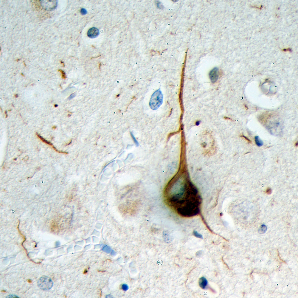
Formalin fixed paraffin embedded section of cerebral cortex of an Alzheimer patient processed with MCA-Ubi-1 using HRP/DAB, giving a brown signal. Also stained with haemotoxylin in blue. A typical flame shaped tangle is seen in a pyramidal neuron in the center and is surrounded by some dystrophic neurites, also strongly ubiquitin positive. Both are commonly seen in cortical and hippocampal Alzheimer brain sections and are typical for this disease, but are rare or absent in healthy brain.
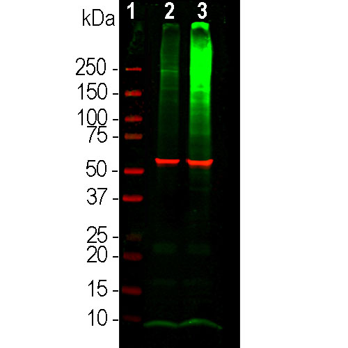
Western blot analysis of HEK293 cell lysates using mouse mAb to ubiquitin, MCA-Ubi-1, dilution 1:1,000 in green. [1] protein standard (red), [2] cells maintained in normal medium, [3] cells treated with 10µM of proteasome inhibitor lactacystin (Lc) for 16hrs. Lysed cells were electrophoresed on 4-20% SDS-PAGE, and transferred to PVDF membranes. The smear detected above the 200kDa standard represent accumulation of ubiquitinated proteins in proteasome inhibitor-Lc treated cells. The prominent band at 8kDa corresponds to monoubiquitin. The same blot was probed with rabbit pAb to HSP60, RPCA-HSP60, dilution 1:5,000 in red, used as a loading control.
Mouse Monoclonal Antibody to Ubiquitin
Cat# MCA-Ubi-1
Price range: $120.00 through $800.00
Ubiquitin is a globular 76 amino acid protein of about 8.5kDa molecular weight which was discovered by biochemical isolation from bovine thymus tissues (1). The protein was found to be highly conserved in amino acid sequence and was detectable in apparently every cell and tissue type, and, being apparently ubiquitously expressed, became known as ubiquitin. Subsequent work showed that ubiquitin has an important role in the targeting of proteins for proteolytic degradation, but has other important functions (2). Proteins to be degraded are covalently coupled to the C-terminus of ubiquitin as a result of the sequential activity of three families of enzymes, the ubiquitin-activating enzymes (E1s), ubiquitin-conjugating enzymes (E2s), and ubiquitin ligases (E3s). While humans express only two E1 enzymes, there are 35 E2 enzymes and hundreds of E3s. The ubiquitinated protein complex may then be degraded in the proteasome. Ubiquitin becomes covalently bonded to many types of pathological inclusions such as the neurofibrillary tangles of Alzheimer’s disease (3), the Lewy bodies of Parkinson’s disease (4), the Pick bodies of Pick’s disease (5) and many others. Ubiquitin can normally be removed from proteins to which it is bound and reused due to the activity of a large family of deubiquitinating enzymes.
This antibody, MCA-Ubi-1, was made in the University of Florida in 1987, and has been continuously marketed since 1989 being sold by EnCor and numerous other vendors. The immunogen was purified bovine blood derived ubiquitin coupled to keyhole limpet hemocyanin with glutaraldehyde (6). The MCA-Ubi-1 is relatively insensitive to formalin fixation and so can be used on paraffin embedded fixed histological sections of human brain for studies of Alzheimer’s and other neurodegenerative diseases. Epitope mapping of MCA-Ubi-1 was performed by generating a series of staggered 20 amino acid peptides which covered the human sequence with 5 amino acid overlap between neighboring peptides. Only the peptide, IQDKEGIPPDQQRLIFAGKQ, amino acids 30-49, inhibited binding of MCA-Ubi-1 to purified bovine ubiquitin, see here. Since the previous and next peptides had no apparent inhibitory effect on antibody binding, the central 10 amino acid segment, GIPPDQQRLI, is likely the most significant component of the MCA-Ubi-1 epitope. This peptide is totally conserved in animals, plants and fungi so MCA-Ubi-1 is applicable to studies of a wide variety of species form human to yeast. MCA-Ubi-1 also works on western blots and can be used to study ubiquitinated proteins which typically accumulate as a high molecular weight smear on western blots if the proteastome is inhibited. We also supply a rabbit polyclonal antibody to ubiquitin, RPCA-Ubi. Mouse select image above left for larger view.

Chromogenic immunostaining of a NBF fixed paraffin embedded human hippocampus section from an Alzheimer’s Disease case. Mouse mAb to ubiquitin, MCA-Ubi-1, dilution 1:2,000, was detected in DAB (brown) following the ImmPress method with citra buffer retrieval. Hematoxylin (blue) was used as the counterstain. The MCA-Ubi-1 antibody strongly labels flame shapped tangles in pyramidal neurons and dystrophic neurites nuclei characteristic of Alzheimer’s disease. This antibody performs well in testing with 4% PFA or standard NBF fixed human and rat tissues. Mouse select image for larger view.

Blots probed with MCA-Ubi-1 of mono and K48 linked polyubiquitin (Boston Biotech, lane 1), monoubiquitin only (2), and 100μg total wet weight of homogenates of rat cerebellum, cortex and brain stem respectively (lanes 3-5). Material was run out on 20% SDS-PAGE and transferred electrophoretically to PVDF. MCA-Ubi-1 binds both mono and polyubiquitin and detects monoubiquitin in cell and tissue lysates.
Additional References;
1. Fortun J, et al. Alterations in degradative pathways and protein aggregation in a neuropathy model based on PMP22 overexpression. Neurobiol Dis. 22:153-164 (2006).
2. Boutajangout A, et al. Characterisation of cytoskeletal abnormalities in mice transgenic for wild-type human tau and familial Alzheimer’s disease mutants of APP and presenilin-1. Neurobiol. Dis. 15:47-60 (2004).
3. Wang DS, et al. Contribution of changes in ubiquitin and myelin basic protein to age-related cognitive decline. Neurosci. Res. 48:93-100 (2004).
4. He CZ, Hays AP. Expression of peripherin in ubiquinated inclusions of amyotrophic lateral sclerosis. J. Neurol. Sci. 217:47-54 (2004).
5. Ungureanu D. et al. Regulation of Jak2 through the ubiquitin-proteasome pathway involves phosphorylation of Jak2 on Y1007 and interaction with SOCS-1. Mol Cell Biol. 22:3316-26 (2002)
6. Wirbelauer C. et al. The F-box protein Skp2 is a ubiquitylation target of a Cul1-based core ubiquitin ligase complex: evidence for a role of Cul1 in the suppression of Skp2 expression in quiescent fibroblasts. EMBO J. 19:5362-75 (2000).
7. Harris KF. et al. Ubiquitin-mediated degradation of active Src tyrosine kinase. Proc Natl Acad Sci U S A. 96:13738-43 (1999).
8. Sternsdorf T, et al. PIC-1/SUMO-1-modified PML-retinoic acid receptor alpha mediates arsenic trioxide-induced apoptosis in acute promyelocytic leukemia. Mol Cell Biol. 19:5170-8 (1999).
9. Marti A, Wirbelauer C, Scheffner M, Krek W. Interaction between ubiquitin-protein ligase SCFSKP2 and E2F-1 underlies the regulation of E2F-1 degradation. Nat Cell Biol. 1:14-9 (1999).
CiteAb link to peer reviewed publications in which this antibody was purchased directly from us, here.
Older peer reviewed publications making use of this antibody.
Perry, G. et al. Proc. Natl. Acad. Sci. USA 84, 3033-3036 (1987)
Shaw, G. and Chau, V. Proc. Natl. Acad. Sci. USA 85, 2854-2858 (1988)
Hirano, S., et al. Cell 70: 293-301 (1992)
Cuervo, A.M., et al. Mol. Biol. 9: 1995-2010 (1995)
Sternsdorf, T., et al. J. Cell Biol. 139: 1621-1634 (1997)
Tae-Wan Kim, et al. J. Biol. Chem. 272: 11006-11010 (1997)
Verdier, F., et al. J. Cell Biol. 273: 28185-28190 (1998)
Laroia, G., et al. Science 284: 499-502 (1999)
Marti, A., et al. Nature Cell Biol. 1: 14-19 (1999)
Sternsdorf, T., et al. Mol. Cell Biol. 19: 5170-5178 (1999)
1. Goldstein G, et al. Isolation of a polypeptide that has lymphocyte-differentiating properties and is probably represented universally in living cells.
PNAS 72:11-5 (1975).
2. Wilkinson K. The discovery of ubiquitin-dependent proteolysis. PNAS 102:15280-2 (2005).
3. Perry G. et al. Ubiquitin is detected in neurofibrillary tangles and senile plaque neurites of Alzheimer disease brains. PNAS 84:3033-6 (1987).
4. Kuzuhara S, et al. Lewy bodies are ubiquitinated. A light and electron microscopic immunocytochemical study. Acta Neuropathol. 75:345-53 (1988).
5. Murayama S. et al. Immunocytochemical and ultrastructural studies of Pick’s disease. Ann. Neurol. 27:394-405 (1990).
6. Shaw G. Chau V. Ubiquitin and microtubule-associated protein tau immunoreactivity each define distinct structures with differing distributions and solubility properties in Alzheimer brain. PNAS 85:2854-8 (1988).
This antibody has been widely used for about 30 years, though mostly sold through our OEM partners. Some of the papers which make use of the antibody supplied by EnCor can be found by searching Google Scholar for “MCA-Ubi AND antibody” or, if you are reading this online, go here.
for a very recent example of the use of MCA-Ubi-1 on western blots to visualize ubiquitin conjugates go to figure 4e in Gladcova C, et al. Mechanism of parkin activation by PINK1. Nature 559:410-414 (2018).
Related products


Recently Viewed Products
-
 Chicken Polyclonal Antibody to FOX3/NeuN
Cat# CPCA-FOX3 Price range: $120.00 through $800.00 -
 Mouse Monoclonal Antibody to Fibrillarin
Cat# MCA-38F3 Price range: $120.00 through $800.00

