| Name: | Mouse monoclonal antibody to Catalase |
| Immunogen: | Full length human catalase expressed in and purified from E. coli. |
| HGNC Name: | CAT |
| UniProt: | P04040 |
| Molecular Weight: | ~60kDa |
| Host: | Mouse |
| Isotype: | IgG1 κ |
| Species Cross-Reactivity: | Human, Rat, Mouse |
| RRID: | AB_3083048 |
| Format: | Purified antibody at 1mg/mL in 50% PBS, 50% glycerol w/v plus 5mM NaN3 |
| Applications: | WB, ICC/IF, IHC |
| Recommended Dilutions: | WB: 1:500. ICC/IF: 1:200, IHC: 1:1,000. |
| Storage: | Shipped on ice. Store at 4°C for short term, for longer term at -20°C. Avoid freeze / thaw cycles. |
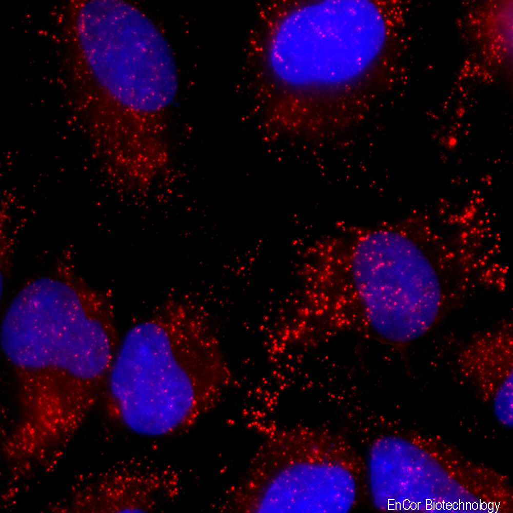
CImmunofluorescent analysis of HeLa cell culture stained with mouse mAb to catalase, MCA-6H14, dilution 1:200 in red. The blue is Hoechst staining of nuclear DNA. MCA-6H14 antibody reveals visicular staining of catalase protein in peroxisomes in the cytoplasm.
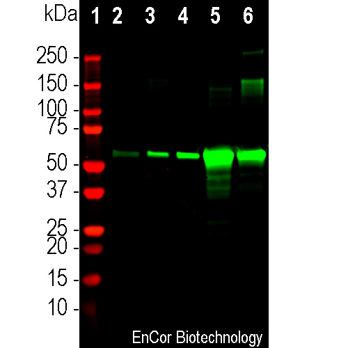
Western blot analysis of mouse different tissue lysates using mouse mAb to Catalase, MCA-6H14, dilution 1:500 in green: [1] protein standard (red), mouse tissue: [2] brain [3] lungs, [4] heart, [5] liver, and [6] kidney. Strong band at about 60kDa corresponds to catalase protein. The protein is particularly abundant in kidney and especially liver.
Mouse Monoclonal Antibody to Catalase
Cat# MCA-6H14
Price range: $250.00 through $1,000.00
Catalase is an enzyme found in all living organisms, including animals, plants, and microorganisms. In eukaryotes it is localized in peroxisomes, which are small, membrane-bound organelles responsible for various metabolic reactions in cells. Catalase catalyses the breakdown of hydrogen peroxide (H2O2) into water (H2O) and oxygen (O2). Hydrogen peroxide is a byproduct of various biochemical reactions in cells and can be harmful if allowed to accumulate, as it can modify and damage cellular proteins, lipids and nucleic acids (1,2). Catalase therefore prevents oxidative stress and maintains cellular health. Catalase forms a tetramer in vivo, and, like hemoglobin and the cytochromes, each subunit contains an iron containing heme group. The enzyme catalase is widely studied in biochemistry as liver is an unusually rich source of the native protein which allowed purification, amino acid sequence determination and crystallization in the 60s and 70s. Catalase activity is important for the regulation of cellular physiology and changes in aging and diseases associated with oxidative stress.
The MCA-6H14 antibody was made against full length recombinant human catalase expressed in and purified from E. coli. The antibody recognizes catalase in human, rodents and many other mammals and is a useful immunocytochemical marker of peroxisomes. It also works well for IHC on neutral buffered formalin fixed and paraffin embedded specimens, see under “additional Info” tab. We also supply a rabbit polyclonal antibody to catalase, RPCA-Catalase. Mouse select image at left for larger view.

Chromogenic immunostaining of a formalin fixed paraffin embedded rat kidney section with mouse mAb to catalase, MCA-6H14, dilution 1:1,000, detected with DAB (brown) using the Vector Labs ImmPRESS method and reagents with citra buffer retrieval. Hematoxylin (blue) was used as the counterstain. The MCA-6H14 antibody strongly labels the peroxisomes in the cytoplasm of kidney tubule cells. This antibody performs well in testing with 4% paraformaldehyde and standard neutral buffered formalin on paraffin sections of fixed mouse, rat, and human tissue. Mouse select image for larger view.

Western blot analysis of different cell line’s lysates using mouse mAb to Catalase, MCA-6H14, dilution 1:500 in green: [1] protein standard (red), [2] HeLa, [3] SH-SY5Y, [4] COS-1, [5] NBL6, [6] A72, [7] NIH/3T3, [8] PC12, and [9] C6. Strong band at about 60kDa corresponds to catalase protein.
1. Kirkman HN. and Gaetani GF. Mammalian catalase: a venerable enzyme with new mysteries. Trends Bioiochem Sci 32:44-50 (2006).
2. Goyal MM and Basak A. Human catalase: looking for complete identity. Protein and Cell 2:888-897 (2010).


