| Name: | Mouse monoclonal antibody to FOX3/NeuN |
| Immunogen: | N-terminal 99 amino acids of human FOX3 expressed in and purified from E. coli |
| HGNC Name: | RBFOX3 |
| UniProt: | A6NFN3 |
| Molecular Weight: | 46, 48kDa |
| Host: | Mouse |
| Isotype: | IgG2b heavy, κ light |
| Species Cross-Reactivity: | Human, Rat, Mouse |
| RRID: | AB_2572267 |
| Format: | Purified antibody at 1mg/mL in 50% PBS, 50% glycerol plus 5mM NaN3 |
| Applications: | WB, IF/ICC, IHC |
| Recommended Dilutions: | WB: 1:1,000 IF/ICC and IHC: 1:1,000-1:2,000 |
| Storage: | Store at 4°C for short term, for longer term at -20°C. |
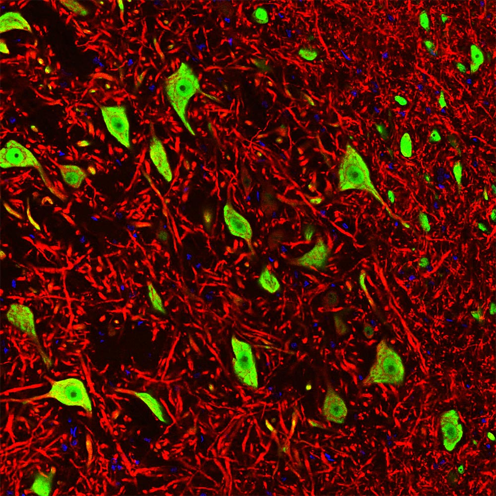
Immunofluorescent analysis of rat brain stem costained with mouse mAb to FOX3/NeuN MCA-1B7 in green, and chicken pAb to microtubule associated protein 2 (MAP2) CPCA-MAP2 in red. Blue is DAPI staining of nuclear DNA. Following transcardial perfusion with 4% paraformaldehyde, the brain was post fixed for 24 hours, cut to 45μM, and free-floating sections were stained with the above antibodies. The FOX3/NeuN antibody selectively stains nuclei and the proximal cytoplasm of neuronal cells while the MAP2 antibody labels dendrites and overlaps with FOX3/NeuN staining in the perikarya of neurons.
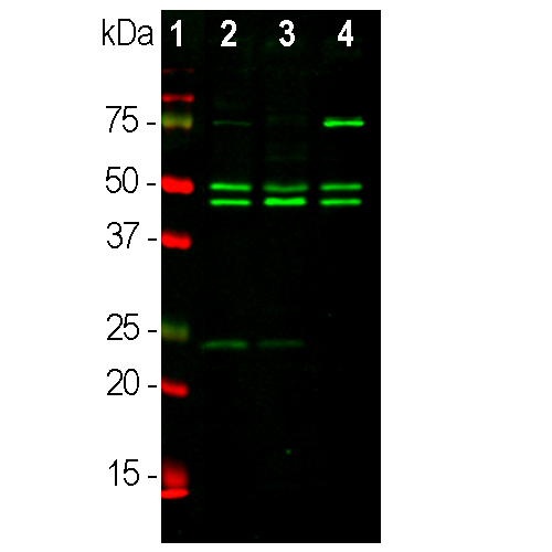
Western blot analysis of whole brain tissue lysates using mouse mAb to FOX3/NeuN MCA-1B7, dilution 1:1,000 in green: [1] protein standard (red), [2] adult rat brain, [3] embryonic E20 rat brain, [4] adult mouse brain. Note the strong twin bands corresponding to the two alternate transcripts of FOX3/NeuN protein with apparent SDS-PAGE molecular weights of 46 and 48kDa. As with other FOX3/NeuN antibodies, an additional band at ~70kDa is revealed in some lysates.
Mouse Monoclonal Antibody to FOX3/NeuN
Cat# MCA-1B7
Price range: $120.00 through $800.00
In the early 90s an unusual protocol resulted in the raising of a mouse monoclonal antibody, clone A60, against a component of neuronal nuclei and proximal perikarya (1). The component was therefore named “NeuN” and was shown to correspond to two protein bands at 46 and 48kDa on western blots. The antibody become very widely used as a reliable neuronal marker, apparently binding to neurons in all vertebrates, and athough a few neuronal cell types were not recognized such as cerebellar Purkinje cells, olfactory mitral cells and many type of retinal neuron, NeuN immunoreactivity has been widely used to identify neurons. The identity of the NeuN protein was unknown until 2009 when Kim et al. (2) showed that it was identical to FOX3, a mammalian homolog of a gene product originally identified in C. elegans and named FOX1 (2,3). There are three mammalian homologs, FOX1, FOX2 and FOX3, which are believed to have a role in the regulation of mRNA splicing (4). All three contain an almost identical central RNA recognition motif or RRM domain, a region of about 90 amino acids found in numerous proteins which in all three molecules specifically binds the hexaribonucleotide UGCAUG (4). Four protein isoforms of FOX3 result from alternate splicing of two exons from the single gene which code for an insert close to the C-terminus and a short C-terminal extension (5). The extension includes a C-terminal proline-tyrosine sequence preceded by hydrophobic amino acids (Φ-PY) which is known to function in nuclear localization, apparently accounting for FOX3 being present in both nuclei and cytoplasm in certain neurons.
The MCA-1B7 antibody was raised against a recombinant human FOX3/NeuN construct based only on the N-terminal sequence, not including the RRM domain and C-terminal regions. The N-terminal regions of FOX1, FOX2 and FOX3 are relatively poorly conserved so we were able to obtain antibodies which recognized FOX3 but not FOX2 or FOX1. The peptide YPPAQYPPPPQNGIPAEYAP, amino acids 5-24, inhibits binding of both MCA-1B7 and the original NeuN antibody to recombinant human FOX3. The central 10 amino acids of the peptide is likely the most significant component of the MCA-1B7 epitope (see here for details). The antibody works well for western blotting and for IF, ICC and IHC (for IHC see data under “Additional Info” tab). The equilibrium dissociation constant (KD) is 2.69 x 10⁻⁹M for MCA-1B7, showing significantly higher affinity than the original Millipore NeuN antibody, which has a KD of 7.95 x 10⁻⁹M under identical conditions. We used the same recombinant immunogen to generate goat and chicken polyclonal antibodies to FOX3/NeuN, GPCA-FOX3, and CPCA-FOX3 respectively. We also generated rabbit polyclonal antibody, RPCA-FOX3, against peptide corresponding to amino acids 5-24 of human FOX3 coupled to KLH. These three antibodies also work in the same way as MCA-1B7 and the original NeuN antibody and are versatile reagents which can be used in double and triple staining protocols and also work well on mouse tissues on which mouse monoclonals present technical problems. Any of our FOX3/NeuN antibodies can be used to quantify the neuron/glial ratio in primary cell cultures and tissue sections from different species (6,7). Mouse select image above left for larger view.
This antibody has become widely used as sold by EnCor and through our numerous OEM partners, and information on this can be viewed using Google scholar by searching for “1B7 AND (NeuN or FOX3) or by selecting here.
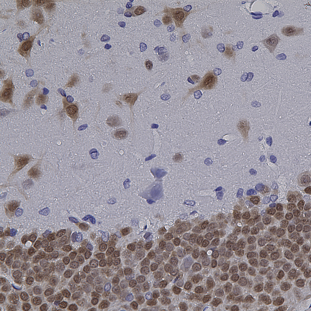
Chromogenic immunostaining of a formalin fixed paraffin embedded rat hippocampus section with mouse mAb to FOX3/NeuN, 1mg/mL MCA-1B7, dilution 1:2,000, detected in DAB (brown) following the Vector Labs ImmPRESS method with citra retrieval. Hematoxylin (blue) was used as the counterstain. This antibody has also been validated on NBF fixed human tissues. Mouse select image for larger view.
We used the data shown below to derive dissociation equilibrium constants (KD values) for specified mouse monoclonal antibodies. The KD is simply the ratio of the dissociation rate (koff) to the association rate (kon). Thus, KD and affinity are inversely related so that the lower the KD value, the higher the affinity of the antibody for its target. The figures below summarize binding data for EnCor’s mouse monoclonal antibody to FOX3/NeuN (MCA-1B7) and Millipore’s mouse monoclonal antibody to the same target (MAB377, clone A60). Experiments were performed in parallel under identical conditions. Note that EnCor’s MCA-1B7 has a significantly higher affinity for FOX3 (KD = 3nM) versus Millipore’s MAB377 (KD = 8nM).

Above: Binding curve set for MCA-1B7 (25nM IgG) and limiting dilutions of recombinant FOX3 peptide (0-500nM) obtained using our in-house label-free bio-layer interferometry system (Octet RED96). Color-coded traces show sensorgram data normalized to baseline after subtraction of 0nM IgG signal from all channels. Traces with overlying fit lines in red indicate their inclusion in the global fit analysis used to derive kinetic parameters listed under the legend (R^2 – goodness of correlation between the fit and data; kon – association rate constant; koff – dissociation rate constant; KD = koff/kon – affinity constant/equilibrium dissociation constant; see EnCor’s validation pipeline for more details). Mouse click on the image to get an enlarged view.

Above: Binding curve set for the Millipore mouse monoclonal antibody MAB377 (25nM IgG) and limiting dilutions of recombinant FOX3 peptide (0-500nM) obtained using our in-house label-free bio-layer interferometry system (Octet RED96). Color-coded traces show sensorgram data normalized to baseline after subtraction of 0nM IgG signal from all channels. Traces with overlying fit lines in red indicate their inclusion in the global fit analysis used to derive kinetic parameters listed under the legend (R^2 – goodness of correlation between the fit and data; kon – association rate constant; koff – dissociation rate constant; KD = koff/kon – affinity constant/equilibrium dissociation constant; see EnCor’s validation pipeline for more details). Mouse click on the image to get an enlarged view.
1. Mullen RJ, Buck CR, Smith AM. NeuN, a neuronal specific nuclear protein in vertebrates. Development 116:201-11 (1994).
2. Hodgkin J, Zellan JD, Albertson DG. Identification of a candidate primary sex determination locus, fox-1, on the X chromosome of Caenorhabditis elegans. Development 120:3681-3689 (1994).
3. Kim KK, Adelstein RS, Kawamoto S. Identification of neuronal nuclei (NeuN) as Fox-3, a new member of the Fox-1 gene family of splicing factors. J. Biol. Chem. 284:31052-61 (2009).
4. Underwood JG, et al. Homologues of the Caenorhabditis elegans Fox-1 protein are neuronal splicing regulators in mammals. Mol. Cell Biol. 25:10005-16 (2005).
5. Dredge BK, Jensen KB. NeuN/Rbfox3 nuclear and cytoplasmic isoforms differentially regulate alternative splicing and nonsense-mediated decay of Rbfox2. PLoS One doi: 10.1371/journal.pone.0021585 (2011).
6. Herculano-Houzel S, Lent R. Isotropic fractionator: a simple, rapid method for the quantification of total cell and neuron numbers in the brain. J. Neurosci. 25:2518-2521 (2005).
7. Azevedo FA. et al. Equal numbers of neuronal and nonneuronal cells make the human brain an isometrically scaled-up primate brain. J. Comp. Neurol. 513:532-41 (2009).
This antibody has become widely used as sold by EnCor and through our numerous OEM partners, and information on this can be viewed using Google scholar by searching for “1B7 AND (NeuN or FOX3) or by selecting here.
Related products


Recently Viewed Products
-
 Mouse Monoclonal Antibody to Neurofilament NF-H
Cat# MCA-AH1 Price range: $120.00 through $800.00 -
 Mouse Monoclonal Antibody to all Actin Isotypes
Cat# MCA-5J11 Price range: $120.00 through $800.00 -
 Mouse Monoclonal Antibody to Neurofilament NF-H
Cat# MCA-9B12 Price range: $120.00 through $800.00

