| Name: | Rabbit Polyclonal Antibody to Neurofilament Light, NF-L-ct |
| Immunogen: | C-terminal peptide of rat NF-L protein with an N-terminal Cys for coupling to KLH |
| HGNC Name: | NEFL |
| UniProt: | P07196 |
| Molecular Weight: | 68-70kDa |
| Host: | Rabbit |
| Isotype: | |
| Species Cross-Reactivity: | Human, Rat, Mouse, Cow, Pig |
| RRID: | AB_2861179 |
| Format: | Affinity purified at 1mg/mL in 50% PBS, 50% Glycerol plus 5mM NaN3 |
| Applications: | WB, IF/ICC, IHC |
| Recommended Dilutions: | WB: 1:10,000-1:20,000. IF/ICC: 1:5,000. IHC:1:5,000 |
| Storage: | Store at 4°C, for longer term, store at -20°C |
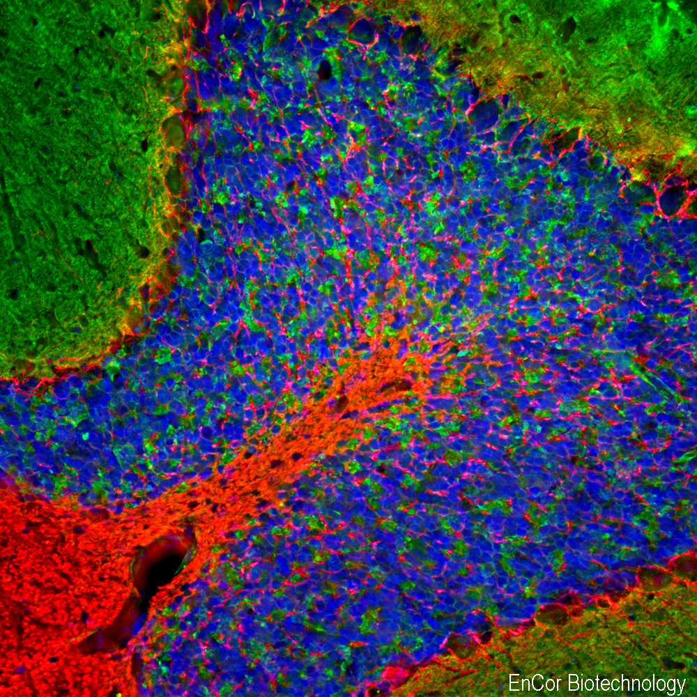
Immunofluorescent analysis of rat cerebellum section stained with rabbit pAb to NF-L RPCA-NF-L-ct, dilution 1:5,000 in red, and costained with mouse mAb to β-synuclein, MCA-6A10, dilution 1:500 in green. The blue is Hoechst staining of nuclear DNA. Following transcardial perfusion with 4% paraformaldehyde, the brain was post fixed for 24 hours, cut to 45µM, and free-floating sections were stained with above antibodies. The NF-L antibody labels dendrites and axons of neuronal cells, and β-synuclein antibody detects protein that is concentrated in synaptic regions.
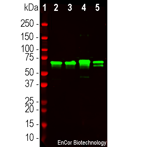
Western blot analysis of tissue lysates using rabbit pAb to NF-L, RPCA-NF-L-ct, dilution 1:20,000, in green. [1] protein molecular weight standard (red), and lysates of rat brain [2], rat spinal cord [3], mouse brain [4] and mouse spinal cord [5]. The strong band at 68kDa corresponds to the NF-L protein.
Rabbit Polyclonal Antibody to NF-L C-terminus
Cat# RPCA-NF-L-ct
Price range: $120.00 through $1,000.00
Neurofilaments are the 10nm or intermediate filament proteins found specifically in neurons, and are composed predominantly of three major proteins called NF-L, NF-M and NF-H, though other filament proteins may be included also. The major function of neurofilaments is likely to control the diameter of large axons (1). NF-L is the neurofilament light or low molecular weight polypeptide and runs on SDS-PAGE gels at 68-70kDa with some variability across species. Antibodies to NF-L are useful for identifying neuronal cells and their processes in cell culture and sectioned material. NF-L antibody can also be useful for the visualization of neurofilament rich accumulations seen in many neurological diseases, such as Lou Gehrig’s disease (ALS), giant axon neuropathy, Charcot-Marie-Tooth disease and others (2-4). Much interest has recently been focused on the detection of NF-L released from neurons into blood and CSF as a surrogate marker of primarily axonal loss in a variety of types of CNS injury and degeneration (5).
RPCA-NF-L-ct was made against amino acids the C-terminal peptide, amino acids 515-543 of rat NF-L in NCBI entry XP_032773476, with an N-terminal cysteine added by which it was coupled to KLH. The antibody binds NF-L from a variety of species including human, rat and mouse and is useful for studies of neurofilament expression and proteolysis. In addition the antibody may be useful for ELISA studies as the epitopic region is known. We recently found that the epitope for this antibody is rapidly degraded during neurodegeneration, see our recent BioRχiv and Brain Communications papers for details. The antibody also works very well on paraffin embedded and formalin fixed human and rodent sections, see data under the “additional info” tag. We market several other NF-L antibodies including rabbit and chicken polyclonals RPCA-NF-L and CPCA-NF-L, both made against full length recombinant human NF-L. We also have several mouse monoclonal antibodies to NF-L including epitope mapped MCA-DA2, MCA-6H63, MCA-1B11 and MCA-1D44. Mouse select image at left for larger view.

Chromogenic immunostaining of a formalin fixed paraffin embedded human cerebellum section with rabbit pAb to NF-L, RPCA-NF-L-ct, dilution 1:5,000, detected with DAB (brown) using the Vector Labs ImmPRESS method and reagents with citra buffer retrieval. Hematoxylin (blue) was used as the counterstain. RPCA-NF-L-ct strongly labels the axons and dendrites of Purkinje cells and the projections of neuronal cells within the granular layer. This antibody performs well in testing with both 4% PFA and standard NBF fixed rat, mouse, and human tissues. Mouse select image for larger view.
This antibody is known to cross react well with human, pig and cow NF-L. As shown below the homologous region of the C-terminus of NF-L is not identical to the rodent immunogen used to make RPCA-NF-L-ct but is substantially similar.
Rat NF-L C-terminus GEE-EDTKESEEEEKKEESAGEEQAAKKKD
Identity GEE E+TKE EEEEKK E AGEEQAAKKKD
Human NF-L C-terminus GEEGEETKEAEEEEKKVEGAGEEQAAKKKD
1. Hoffman et al. Neurofilament gene expression:a major determinant of axonal caliber. PNAS 84:3472-6 (1987).
2. Perrot R, et al. Review of the Multiple Aspects of Neurofilament Functions, and their Possible Contribution to Neurodegeneration. Mol. Neurobiol. 38:27-65 (2008).
3. Lépinoux-Chambaud C. Eyer J. Review on intermediate filaments of the nervous system and their pathological alterations. Histochem. Cell Biol. 140:13-22 (2013).
4. Liu Q. et al. Neurofilamentopathy in Neurodegenerative Diseases. Open Neurol. J. 5:58–62 (2011).
5. Bacioglu M, et al. Neurofilament light chain in blood and CSF as marker of disease progression in mouse models and in neurodegenerative diseases. Neuron 91:56-66 (2016).
Related products


Recently Viewed Products
-
 Rabbit Polyclonal Antibody to c-FOS
Cat# RPCA-c-FOS Price range: $150.00 through $1,000.00 -
 Rabbit Polyclonal Antibody to ALDH1L1
Cat# RPCA-ALDH1L1 Price range: $120.00 through $800.00

