| Name: | Rabbit polyclonal antibody to MAP2D |
| Immunogen: | Full length recombinant human MAP2D protein Prot-r-MAP2D expressed in and purified from E. coli. |
| HGNC Name: | MAP2 |
| UniProt: | P11137 |
| Molecular Weight: | MAP2A/B ~280 kDa, MAP2C/D ~70kDa by SDS-PAGE |
| Host: | Rabbit |
| Isotype: | |
| Species Cross-Reactivity: | Human, Rat, Mouse |
| RRID: | AB_3083051 |
| Format: | Purified antibody at 1mg/mL in 50% PBS, 50% glycerol plus 5mM NaN3 |
| Applications: | WB, IF/ICC, IHC |
| Recommended Dilutions: | WB: 1:10,000-20,000. IF/ICC 1:2,000-5,000. IHC: 1:4,000 |
| Storage: | Stable at 4°C for one year, for longer term store at -20°C |
Immunofluorescent analysis of a section of cerebellum from an adult rat stained with rabbit pAb to Microtubule Associated Protein 2D (MAP2D), RPCA-MAP2D, dilution 1:2,000 in green, and costained with mouse mAb to Glial Fibrillary Acidic Protein (GFAP), MCA-5C10, dilution 1:1,000 in red. The blue is Hoechst staining of nuclear DNA. The MAP2 antibody stains dendrites and perikarya of neurons, while the GFAP antibody labels network of astrocytic glial cells.
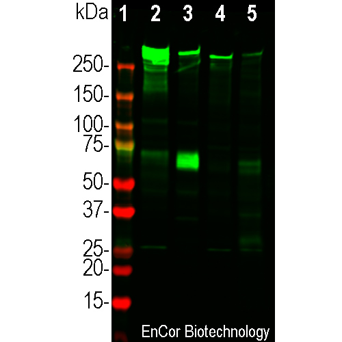
Western blot analysis of different tissue lysates using rabbit pAb to Microtubule Associated Protein 2D (MAP2D), RPCA-MAP2D, dilution 1:20,000 in green: [1] protein standard (red) and extracts of [2] adult rat brain, [3] E20 rat brain, [4] adult mouse brain, and [5] cow cerebellum. Strong band at about 280 kDa mark corresponds to two major high molecular isoforms of MAP2 protein referred to as MAP2A and MAP2B. The band at about 70kDa represents the MAP2D isotype detected exclusively in the embryonic developing brain, which in later development is replaced by MAP2A and MAP2B.
Rabbit Polyclonal Antibody to MAP2D
RPCA-MAP2D
Price range: $300.00 through $1,200.00
Microtubules are 25nm diameter protein rods found in most eukaryotic cells and are associated with a family of proteins called microtubule associated proteins (MAPs). MAPs play a crucial role in the regulation of microtubule dynamics and interactions in vivo. MAP2 was originally named as one of the higher molecular weight MAPs with an SDS-PAGE molecular weight of about 280kDa (1-3). There is a single mammalian MAP2 gene which may generates two high molecular weight proteins of ~280kDa on SDS-PAGE named MAP2A and MAP2B and multiple lower molecular weight forms usually named MAP2C and MAP2D which run on SDS-PAGE gels at 60-70kDa. The lower molecular weight forms are found in neurons early in development, but as the animal matures they are replaced by the higher molecular weight forms (1,2). MAP2 isoforms are expressed only in neurons in perikarya and dendrites, so MAP2 antibodies are useful for identifying neurons in cell culture and sectioned material (4-8). MAP2C and D contain an “intrinsically unstructured region”, one of the prototypes for this widespread type of protein sequence (9). Since MAP2C and D are expressed earlier in development than MAP2A and B this antibody can be used for monitoring early neuronal development, though it is also useful as a general marker for neurons and dendrites in mature tissues.
This antibody was made against a recombinant full length form of human MAP2D and was found, as expected, to bind all four MAP2 gene products since it binds to the shared core region of all these molecules. The antibody will therefore recognize both the low molecular weight forms of MAP2 seen in development as well as the larger forms seen in the mature CNS. As shown here, the antibody works well for western blotting, IF, ICC and IHC. EnCor markets mouse monoclonal antibodies specific for MAP2A and MAP2B MCA-4H5 and MCA-5H11. EnCor also markets chicken and goat polyclonal antibodies to MAP2, CPCA-MAP2 and GPCA-MAP2. Mouse select image above left for larger view.

Chromogenic immunostaining of a 4% paraformaldehyde fixed paraffin embedded rat cerebellum section with rabbit pAb to MAP2D, RPCA- MAP2D, dilution 1:4,000, detected with DAB (brown) using the Vector Elite ABC-HRP detection and reagents with citra buffer retrieval. Hematoxylin (blue) was used as the counterstain. RPCA-MAP2D strongly labels Purkinje cell perikarya and dendrites. This antibody performs well in testing with both 4% PFA and standard neutral buffer formalin fixed and paraffin embedded rat, mouse and human tissues. Mouse select image for larger view.

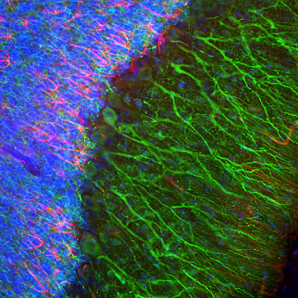
Immunofluorescent analysis of a section of adult rat hippocampus stained with rabbit pAb to Microtubule Associated Protein 2D (MAP2D), RPCA-MAP2D, dilution 1:2,000 in green, and with mouse mAb to Glial Fibrillary Acidic Protein (GFAP), MCA-5C10, dilution 1:1,000 in red. The blue is Hoechst staining of nuclear DNA. The MAP2D antibody stains dendrites and perikarya of neurons, while the GFAP antibody labels network of astrocytic glial cells. Mouse select image above left for larger view.
1. Dehmelt H, Halpain S. The MAP2/Tau family of microtubule-associated proteins.
Genome Biol. 6:204 (2005).
2. Nunez J. Immature and mature variants of MAP2 and tau proteins and neuronal plasticity. Trends Neurosci. 11:477-9 (1998).
3. Vallee R. A taxol-dependent procedure for the isolation of microtubules and microtubule-associated proteins (MAPs). J. Cell Biol. 92:435-42 (1992).
4. Goetz AK, et al. Temporally restricted substrate interactions direct fate and specification of neural precursors derived from embryonic stem cells. PNAS 103:11063-8 (2006).
5. Walton NM et al. Gliotypic neural stem cells transiently adopt tumorigenic properties during normal differentiation. Stem Cells 27:280-9 (2009).
6. Gasser A, et al. An ankyrinG-binding motif is necessary and sufficient for targeting Nav1.6 sodium channels to axon initial segments and nodes of Ranvier. J. Neurosci. 32:7232-43 (2012).
7. Rush AM, et al. Differential modulation of sodium channel Nav1.6 by two members of the fibroblast growth factor homologous factor 2 subfamily. Eur. J. Neurosci. 23:2551-62 (2006).
8. Eckenstein FP, McGovern T, Kern D, Deignan J. Neuronal vulnerability in transgenic mice expressing an inducible dominant-negative FGF receptor. Exp. Neurol. 198:338-49 (2006).


