| Name: | Rabbit Polyclonal Antibody to Coronin 1a |
| Immunogen: | C-terminal peptide of human coronin 1a couple to KLH |
| HGNC Name: | CORO1A |
| UniProt: | P31146 |
| Molecular Weight: | ~55kDa |
| Host: | Rabbit |
| Isotype: | |
| Species Cross-Reactivity: | Human, Rat, Mouse, Cow, Pig |
| RRID: | AB_2229659 |
| Format: | Aliquot of serum plus 5mM NaN3 |
| Applications: | WB, IF/ICC |
| Recommended Dilutions: | WB: 1:5,000, IF/ICC: 1:500-1:1,000 |
| Storage: | Store at 4°C for short term, for longer term at -20°C |
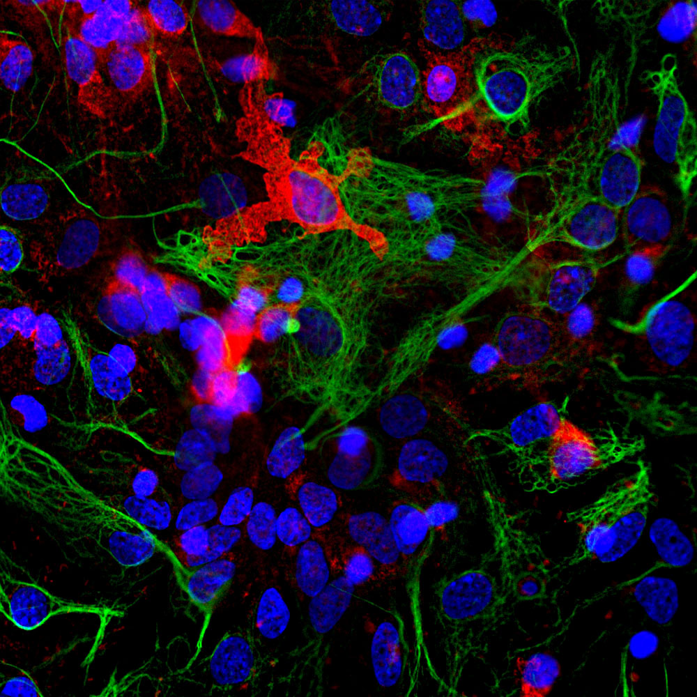
Immunofluorescent analysis of cortical neuron-glial cell culture from E20 rat stained with rabbit pAb to coronin 1a, RPCA-Cor1a, dilution 1:1,000 in red, and costained with mouse mAb to GFAP, MCA-5C10, dilution 1:1,000 in green. The blue is Hoechst staining of nuclear DNA. The coronin 1a antibody labels protein expressed in the cytoplasm of microglia cells, while GFAP antibody stains intermediate filaments in astrocytic cells.
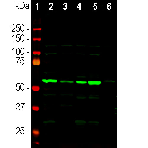
Western blot analysis of tissue lysates using rabbit pAb to coronin 1a, RPCA-Cor1a, dilution 1:5,000: [1] protein standard, [2] mouse brain, [3] rat brain, [4] cow cerebellum, [5] cow cortex, and [6] pig spinal cord. The strong single band about 55kDa corresponds to the coronin 1a protein.
Rabbit Polyclonal Antibody to Coronin 1a
Cat# RPCA-Cor1a
Price range: $120.00 through $800.00
Coronin was originally discovered in the slime mold Dictyostelium, where it was found to be involved in the chemotactic response of these amoeboid cells (1,2). The name derives from from “corona”, which is Latin for crown, since the protein was localized at the leading edge or crown of these highly motile cells. In mammals, there are at least five coronin family proteins derived from 5 distinct genes. These are named coronins 1 to 5 in one nomenclature or coronins 1a and 1b, 2a, 2b and 2c in another (1,2). Coronins are members of the WD or WD40 family of proteins, which are generally involved in mediating important protein to protein interactions (3). The basic structure of WD molecules is a seven bladed β-propellor structure each blade being based on some variant of a ~40 amino acid WD (Trp-Asp) containing peptide. Structural studies show that coronin 1a is a typical 7 bladed β-propellor member of this protein family (4) Coronin 1a, a.k.a coronin 1 is found exclusively in hematopoietic lineage cells such as lymphocytes, macrophages and neutrophils. The only hematopoietic cells normally found within the neuropil and white matter of the central nervous system are the microglia, so antibodies to coronin 1a are useful to identify this important cell type (5). Microglia mediate immune responses in the CNS and are able to actively phagocytose infectious or damaged material. They are very active cells, constantly migrating through the brain and interacting with other cells (6). A typical response to CNS damage or disease is increase the number of microglia at the compromised site.
The RPCA-Cor1a antibody was made against the C-terminal peptide of human coronin 1a chemically coupled to keyhole limpet hemocyanin carrier protein. The antibody is known to work on many mammalian species both on western blots and on cells grown in tissue culture. EnCor markets another antibody which is useful in the identification of microglia which binds a peptide in IBA1, a.k.a AIF1, RPCA-IBA1. Like coronin 1a, IBA1 is a protein expressed only in hematopoietic cells and so is also a useful marker of microglia. Mouse select image at left for a larger view.

Chromogenic immunostaining of a formalin fixed paraffin embedded human brain cortex section with rabbit pAb to coronin 1a, RPCA-Cor1a, dilution 1:2,000, detected with DAB (brown) using the Vector Labs ImmPRESS method and reagents with citra buffer retrieval. Hematoxylin (blue) was used as the counterstain. The RPCA-Cor1a antibody specifically labels the perikarya and processes of microglial cells. This antibody performs well in testing with 4% PFA and standard NBF fixed mouse, rat, and human tissue. Mouse select image for larger view.
1. de Hostos E. The coronin family of actin-associated proteins. Trends Cell Biol. 9:345-50 (1999).
2. Rybaki V. Clemen CS. Coronin proteins as multifunctional regulators of the cytoskeleton and membrane trafficking. BioEssays 27:625-32 (2005).
3. Smith TF. et al. The WD repeat: a common architecture for diverse functions. Trends Biochem. Sci. 24:181-5 (1999).
4. Appleton BA. Wu P. Weisman C. The crystal structure of murine coronin-1: a regulator of actin cytoskeletal dynamics in lymphocytes. Structure 14:87-96 (2006).
5. Ahmed Z, et al. The Actin Binding Proteins Coronin-1a and IBA-1 Are Effective Microglial Markers for Immunohistochemistry. J. Histochem. Cytochem. 55:687-700 (2007).
6. Nimmerjahn A, Kirchhoff F, Helmchen F. Resting Microglial Cells Are Highly Dynamic Surveillants of Brain Parenchyma in Vivo. Science 308:1314-8 (2005).
Related products


Recently Viewed Products
-
 Poster #09
Price range: $5.00 through $50.00
Poster #09
Price range: $5.00 through $50.00
-
 Mouse Monoclonal Antibody to GFAP
Cat# MCA-5C10 Price range: $120.00 through $800.00 -
 Mouse Monoclonal Antibody to Neurofilament NF-H
Cat# MCA-AH1 Price range: $120.00 through $800.00

