| Name: | Rabbit polyclonal antibody to c-FOS |
| Immunogen: | Full length recombinant human protein expressed in and purified from E. coli. |
| HGNC Name: | FOS |
| UniProt: | P01100 |
| Molecular Weight: | 50-65kDa by SDS-PAGE |
| Host: | Rabbit |
| Isotype: | |
| Species Cross-Reactivity: | Human, rat, mouse |
| RRID: | AB_2572236 |
| Format: | Affinity purified antibody at 1mg/mL in 50% PBS, 50% glycerol plus 5mM NaN3 |
| Applications: | WB, IF/ICC, IHC |
| Recommended Dilutions: | WB: 1:2,000 IF/ICC: 1:2,000 IHC: 1:5,000-10,000 |
| Storage: | Store at 4°C for short term, for longer term at -20°C |
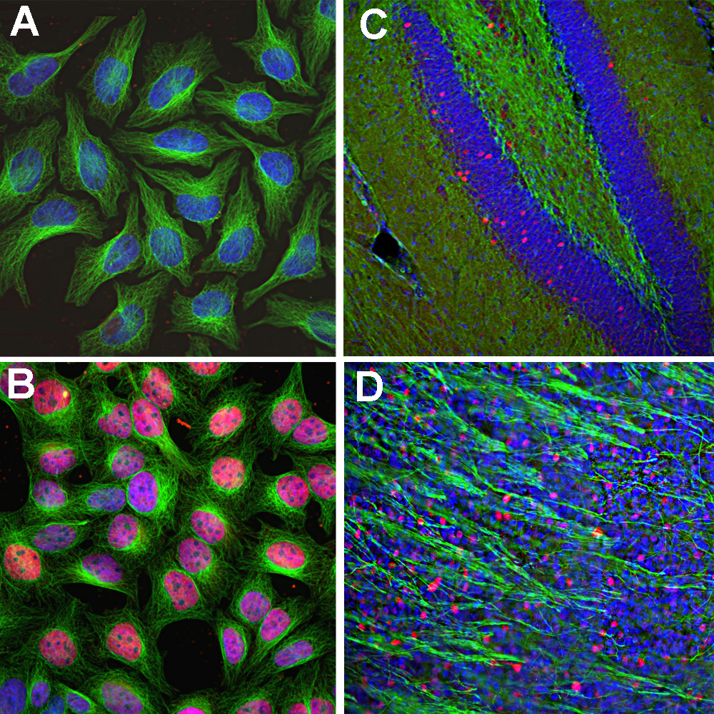
Immunofluorescent analysis of HeLa cells stained with rabbit pAb to c-FOS, RPCA-c-FOS, dilution 1:2,000 in red, and mouse mAb to tubulin, MCA-1B12, dilution 1:10,000, in green. The blue is DAPI staining of nuclear DNA. HeLa cells were kept in fetal bovine serum (FBS) – free media for 36 hours. Then the cells were treated with PBS (A), as a control, or stimulated with 20% FBS (B) for 30 min. c-FOS antibody labels only the nuclei of stimulated cells. Mouse hippocampus (C) or olfactory bulb sections (D) stained with RPCA-c-FOS, dilution 1:10,000 in red, and mouse mAb to NF-L, MCA-7D1, dilution 1:5,000, in green. The blue is Hoechst staining of nuclear DNA. Following transcardial perfusion of mouse with 4% paraformaldehyde, brain was post fixed for 24 hours, cut to 45μM, and free-floating sections were stained with above antibodies. The c-FOS antibody stains only nuclei of spontaneously active neurons. NF-L is expressed in axons of neuronal cells.
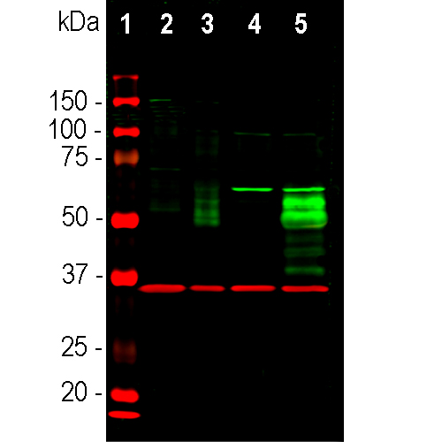
Western blot analysis of cell lysates using rabbit pAb to c-FOS, RPCA-c-FOS, dilution 1:2,000, in green, and mouse mAb to GAPDH, MCA-1D4, dilution 1:5,000, in red, used as a loading control. [1] protein standard (red), [2] HeLa cells grown in FBS free media, [3] HeLa cells stimulated with 20% FBS for 2 hours after being in FBS free media for 36 hours, [4] rat cortical neurons, [5] rat cortical neurons treated with membrane depolarization buffer for 5 hours. Multiple bands at 50-65 kDa in stimulated or treated cell lysates, correspond to different isoforms of the c-FOS protein.
Rabbit Polyclonal Antibody to c-FOS
Cat# RPCA-c-FOS
Price range: $150.00 through $1,000.00
The FOS gene and protein were originally identified as the transforming element in a viral oncogene. The transforming protein was named v-FOS, for viral FOS, and the normal cellular non-transforming proto-oncogene was called c-FOS, for cellular FOS. FOS is an acrynym for “FBJ murine osteogenic sarcoma”, the virus in which the gene product was first discovered. The c-FOS protein is a normal gene acting as an on/off switch controlling the expression of many other genes. The v-FOS form is mutated to stay in the on position, this persistently activating other genes and promoting unregulated cell division. The unmutated c-FOS is an “immediate-early” gene, so-called because protein expression is usually very low but increases rapidly and transiently in response to a wide array of stimuli including serum, growth factors, tumor promoters, cytokines, and UV radiation. Newly expressed c-FOS protein associates with JUN family and other basic leucine-zipper (bZIP) proteins to create a variety of activator protein-1 (AP-1) complexes (1). AP-1 complexes specifically activate the expression of many other genes and so regulate cellular responses to stimuli which may result in cell proliferation, differentiation, neoplastic transformation, apoptosis, and response to stress (2). The regulated expression of c-FOS therefore plays an important role in many cellular functions. Site specific phosphorylation activates c-FOS, while sumoylation of c-FOS inhibits the AP-1 transcriptional activity (3,4). Since c-FOS expression is induced in neurons which are rapidly firing action potentials, appropriate c-Fos antibodies can be used to identify activated neurons in tissues (5). Using current techniques it is possible to follow processes of such cells or obtain data on their mRNA expression.
The RPCA-c-FOS antibody was made against recombinant full length human c-FOS expressed in and purified from E. coli. It can be used to identify activated cells in cell culture and in formalin fixed floating and paraffin embedded sections and to follow c-FOS expression in western blots of cell and tissue homogenates. The same immunogen was used to generate a mouse monoclonal antibody to c-FOS, MCA-2H2, an antibody with similar properties. Mouse select image at left for larger view.

Chromogenic immunostaining of a 4% PFA fixed paraffin embedded rat hippocampus section with rabbit pAb to c-FOS, RPCA-c-FOS, dilution 1:5,000, detected with DAB (brown) using the Vector Labs ImmPRESS method and reagents with citra buffer retrieval. Hematoxylin (blue) was used as the counterstain. The RPCA-c-FOS antibody specifically labels activated neurons. This antibody performs well in testing with 4% PFA and NBF fixed mouse, human, and rat tissues. Mouse select image for larger view.
This antibody has been shown to work very on cells grown in tissue culture, being absent from serum starved cells but heavily upregulated following serum addition. See http://encorbio.com/Data/cFosabpics.html for more details. Similar finding were made with the EnCor monoclonal antibody to c-FOS, MCA-2H2.
Additional References
6. Bossis G, Malnou CE, Farras R, Andermarcher E, Hipskind R, Rodriguez M, Schmidt D, Muller S, Jariel-Encontre I, Piechaczyk M. Down-regulation of c-Fos/c-Jun AP-1 dimer activity by sumoylation. Mol. Cell Biol.25:6964-79 (2005).
7. Day HE, Kryskow EM, Nyhuis TJ, Herlihy L, Campeau S. Conditioned Fear Inhibits c-fos mRNA Expression in the Central Extended Amygdala. Brain Res. 1229:137–46 (2008).
8. Hoffman G, Smith MS, Verbalis JG. c-Fos and related immediate early gene products as markers of activity in neuroendocrine systems. Fronties in Neuroendocrinology 14:173-213 (1993).
9. Van Elzakker M, Fevurly RD, Breindel T, Spencer RL. Environmental novelty is associated with a selective increase in Fos expression in the output elements of the hippocampal formation and the perirhinal cortex. Learn. Mem. 15:899–908 (2008).
10. Dragunow M, Faull R. The use of c-fos as a metabolic marker in neuronal pathway tracing. J. of Neurosci. Mets. 29:261–5 (1989).
1. Mildle-Langosch K. The Fos family of transcription factors and their role in tumourigenesis. Eur. J. Cancer 41:2449-2461 (2005)..
2. Chiu R, et al. The c-Fos protein interacts with c-Jun/AP-1 to stimulate transcription of AP-1 responsive genes. Cell 54:541–52 (1988).
3. Karin M. The regulation of AP-1 activity by mitogen activated protein kinases. J Biol Chem. 270:16483-6 (1995).
4. Bossis G, et al. Down-regulation of c-Fos/c-Jun AP-1 dimer activity by sumoylation. Mol Cell Biol.25(16):6964-79 (2005).
5. Dragunow M, Faull R. The use of c-fos as a metabolic marker in neuronal pathway tracing. J. Neurosci. Mets. 29:261–265 (1989).
This is a new antibody but peer reviewed publications using it are beginning to come on line. So if you perform a Google Scholar search for “MCA-2H2” several publications will appear, or you can just select here.
Related products


Recently Viewed Products
-
 Rabbit Polyclonal Antibody to ALDH1L1
Cat# RPCA-ALDH1L1 Price range: $120.00 through $800.00

