| Name: | Protein standard for Uman type NF-L assays |
| HGNC Name: | NEFL |
| RRID: | NA |
| Format: | 0.5mg/mL in 6M Urea and phosphate buffer at pH=7.4 |
| Applications: | ELISA standard, immunogen |
| Storage: | Store at -20°C |
| Uniprot: | P07196 |
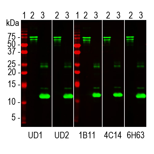
Western blots of Uman NF-LIGHT™ antibodies and a set of EnCor reagents on PROT-r-NF-L and PROT-r-NF-L-Stan. Lanes labelled 1 in red are protein standards of indicated molecular weights. Lanes labelled 2 were loaded with full length recombinant human NF-L, PROT-r-NF-L, while lanes labelled 3 were loaded with PROT-r-NF-L-Stan. The full length protein runs at about 75kDa, while PROT-r-NF-L-Stan runs at about 12kDa. All five antibodies recognize both constructs. UD1 is also known as 2.1 is the detection reagent in the Uman NF-LIGHT™ assay while UD2, also known as 47.3 is the capture reagent. The three other lanes show results obtained with EnCor antibodies MCA-1B11, MCA-4C14 and MCA-6H63 respectively as indicated.
Recombinant Human NF-L Standard
Cat# PROT-r-NF-L-Stan
Price range: $300.00 through $2,000.00
Neurofilaments are the 10nm or intermediate filament proteins found specifically in neurons, and are composed predominantly of four major proteins called NF-L, NF-M, NF-H and α-internexin. NF-L, NF-M and NF-H were named based on their apparent molecular weight on SDS-PAGE gels, so NF-L is low or light, NF-M is medium or middle and NF-H is high or heavy. On SDS-PAGE NF-L runs at 68-70kDa, NF-M at 145-160kDa and NF-H at 200-220kDa with some species variability, larger species tending to have larger molecules. These three proteins are major components of large diameter axons in the adult, while α-internexin is a more major component of the developing nervous system, although still present in the adult. NF-L and other neurofilament subunits accumulate in many neurological diseases and mutations in the protein coding region of the human NF-L gene cause some forms of Charcot-Marie-Tooth disease (2-4). NF-L is a very abundant protein particularly concentrated in large diameter axons and may leak into blood and CSF following various kinds of axonal injury and/or degeneration. There has therefore been much recent interest in the detection of NF-L in CSF and blood as a surrogate marker of neuronal damage and degeneration (5).
A codon optimized cDNA designed to express amino acids 306-364 of human neurofilament NF-L was inserted into pET29a(+) eukaryotic expression vector, which adds a C-terminal in frame His-tag and some other vector derived sequence. We recently showed that both epitopes for the antibodies used in the Uman NF-LIGHT™ and Quanterix Simoa™ NF-L assays (6) bind to this region of NF-L, so this protein will be an excellent standard for assays of this type (7 or download our BioRχiv preprint). We included two tryptophan residues to allow accurate spectrophotometric quantification. The construct was transformed into E. coli and purified in 6M urea using immobilized metal affinity chromatography. Purified protein was diluted to 0.5mg/mL and is supplied in 6M urea.
Human NF-L sequence was based on that was NP_006149.2 which was inserted into the eukaryotic expression vector pET29a(+) which adds an C terminal His-tag and some other sequence, shown in red below. The sequence in black is amino acids 306-364, the region of human NF-L including the epitopes for both monoclonal antibodies employed in the Uman NF-LIGHT™ and Simoa™ assays and also our novel antibodies raised against this region. The sequence also includes two tryptophan residues, shown in blue, which were added to ensure accurate spectrophotometric quantification at a wavelength of 280nm.
MKETAAAKFE RQHMDSPDLG TLVPRGSMAD IGSEFWSESR RLLKAKTLEI EACRGMNEAL 60
EKQLQELEDK QNADISAMQD TINKLENELR TTKSEWVDKL AAALEHHHHH H 111
Number of amino acids: 111
Molecular weight: 12705.26
Theoretical pI: 5.60
Amino acid composition:
Ala (A) 12 10.8%
Arg (R) 6 5.4%
Asn (N) 4 3.6%
Asp (D) 7 6.3%
Cys (C) 1 0.9%
Gln (Q) 5 4.5%
Glu (E) 14 12.6%
Gly (G) 4 3.6%
His (H) 7 6.3%
Ile (I) 4 3.6%
Leu (L) 12 10.8%
Lys (K) 9 8.1%
Met (M) 5 4.5%
Phe (F) 2 1.8%
Pro (P) 2 1.8%
Ser (S) 7 6.3%
Thr (T) 6 5.4%
Trp (W) 2 1.8%
Tyr (Y) 0 0.0%
Val (V) 2 1.8%
Total number of negatively charged residues (Asp + Glu): 21
Total number of positively charged residues (Arg + Lys): 15
Extinction coefficients are in units of M-1 cm-1, at 280 nm measured in water.
Ext. coefficient 11000
Abs 0.1% (=1 g/l) 0.866, assuming all pairs of Cys residues form cystines
Ext. coefficient 11000
Abs 0.1% (=1 g/l) 0.866, assuming all Cys residues are reduced
1. Hoffman et al. Neurofilament gene expression: a major determinant of axonal caliber. PNAS 84:3472-6 (1987).
2. Perrot R, et al. Review of the Multiple Aspects of Neurofilament Functions, and their Possible Contribution to Neurodegeneration.Mol. Neurobiol. 38:27-65 (2008).
3. Lépinoux-Chambaud C. Eyer J. Review on intermediate filaments of the nervous system and their pathological alterations. Histochem. Cell Biol. 140:13-22 (2013).
4. Liu Q. et al. Neurofilamentopathy in Neurodegenerative Diseases. Open Neurol. J. 5:58–62 (2011).
5. Bacioglu M, et al. Neurofilament light chain in blood and CSF as marker of disease progression in mouse models and in neurodegenerative diseases. Neuron 91:56-66 (2016).
6. Norgren N, et al. Monoclonal antibodies selective for low molecular weight neurofilaments. Hybrid. Hybridomics 21:53-59 (2002).
7. Shaw et al. Uman Type NF-L Antibodies Are Effective Reagents for the Imaging of Neurodegeneration. BioRχiv DOI 10.1101/2022.08.27.504533v1 2022.


