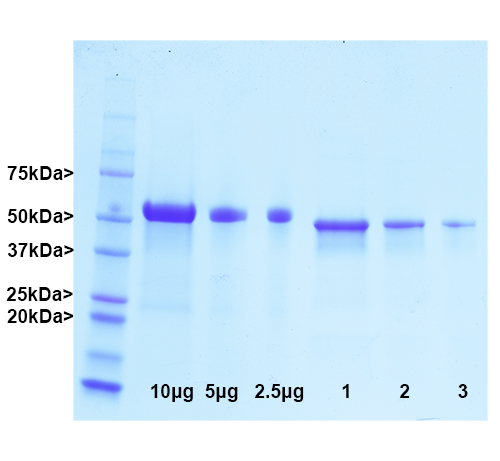
Pig GFAP was purified by a modification of the method of Leung and Liem (1). Cytoskeletal material was prepared by homogenization, detergent extraction and centrifugation to produce a pellet containing intermediate filaments and other stable protein complexes. This material was dissolved in 6M urea and separated by ion exchange chromatography on hydroxyapatite and then on a phosphate gradient on DEAE-cellulose to produce pure GFAP. The gel shows molecular weight standards in lane S and the indicated amounts of BSA in the next three lanes. The last three lanes show three loadings of the GFAP fraction.
Native Pig GFAP Protein
Cat# Prot-m-GFAP
Price range: $300.00 through $2,000.00
Glial Fibrillary Acidic Protein (GFAP) is a major protein of the nervous system and is localized in astrocytes, stem cells, Bergmann glia and non-myelinating Schwann cells. It may also be found in retinal Mueller cells in pathological states, and the levels of the protein generally increase in damage and disease states. GFAP assembles to form 10nm or intermediate filaments in the cytoplasm, and these filaments appear to have an important structural role in the cell. Recent work suggests that measurement of the levels of GFAP in blood and CSF gives information about CNS damage and disease states. The Prot-m-GFAP product was purified from pig spinal cords using biochemical methods and can be used as an ELISA standard or to generate GFAP antibodies. Recombinant forms of GFAP are also available based on the human (Prot-r-GFAP) and rat sequences Prot-r-GFAP-rat.
| Name: | Purified porcine GFAP |
| Immunogen: | Pig GFAP isolated from spinal cord |
| HGNC Name: | GFAP |
| UniProt: | |
| Molecular Weight: | 50kDa |
| Host: | Pig |
| Isotype: | |
| Species Cross-Reactivity: | NA |
| RRID: | Pending |
| Format: | 1mg/mL in 6M Urea |
| Applications: | ELISA, western blotting standard |
| Recommended Dilutions: | |
| Storage: | Store at -20°C |
Glial Fibrillary Acidic Protein (GFAP) is a major protein of the nervous system and is localized in astrocytes, stem cells, Bergmann glia and non-myelinating Schwann cells. It may also be found in retinal Mueller cells in pathological states, and the levels of the protein generally increase in damage and disease states. GFAP assembles to form 10nm or intermediate filaments in the cytoplasm, and these filaments appear to have an important structural role in the cell.
Glial Fibrillary Acidic Protein (GFAP) was discovered by Amico Bignami and coworkers as a major fibrous protein of multiple sclerosis plaques (2). It was subsequently found to be a member of the 10nm or intermediate filament protein family, specifically the intermediate filament protein family Class III, which also includes peripherin, desmin and vimentin. The one mammalian GFAP gene produces several transcripts producing several different possible protein products (1). In pigs there are two major protein products, one of 428 amino acids and a second including a 40 amino acid insert in the C-terminal region. The two proteins are of about equal abundance and run on gels at about 50kDa and 54kDa, so purified pig GFAP therefore presents as two closely spaced bands as shown in the gel image. The larger protein is in Genbank here and the lower here. Both forms bind most available GFAP antibodies including our mouse monoclonal MCA-5C10 and our rabbit and chicken polyclonals, RPCA-GFAP and CPCA-GFAP. GFAP is strongly and specifically expressed in astrocytes and certain other astroglia in the central nervous system, in satellite cells in peripheral ganglia, and in non-myelinating Schwann cells in peripheral nerves. In many damage and disease states GFAP expression is heavily upregulated in astrocytes. In addition neural stem cells frequently strongly express GFAP. Antibodies to GFAP are therefore very useful as markers of astrocytic cells and neural stem cells. In addition many types of brain tumor, presumably derived from astrocytic cells, heavily express GFAP. Alexander’s disease was recently shown to be caused by point mutations in the protein coding region of the GFAP gene (3). All forms of Alexander disease are characterized by the presence of Rosenthal fibers, which are GFAP containing cytoplasmic inclusions found in astrocytes. There has been considerable recent interest in GFAP due to potential use as a damage and degeneration biomarker, since it can be detected in blood and CSF following various kinds of CNS damage and disease states (5). This GFAP preparation is an excellent protein standard for such experiments. Since the human and rodent proteins are somewhat different in primary sequence from each other and from the pig protein, we have also generated recombinant forms of these specifically Prot-r-GFAP and Prot-r-GFAP-rat. The HGNC name for this protein is GFAP.
1. Leung, C. L. and Liem, R. K. H. Isolation of intermediate filaments. Curr. Prot. Cell Biol. 3:Unit 3.23 doi: 10.1002/0471143030.cb0323s31 (2006).
2. Bignami A, Eng LF, Dahl D, Uyeda CT. Localization of the glial fibrillary acidic protein in astrocytes by immunofluorescence. Brain Res. 43:429-35 1972.
3. Brenner M. et al.. Mutations in GFAP, encoding glial fibrillary acidic protein, are associated with Alexander disease. Nat Genet 27:117-20 2001
4. Liem RKH, Yen SH, Salomon GD and Shelanski ML. Intermediate filaments in nervous tissues. J Cell Biol 79:637-745 (1978).
5. Moeton, M. et al. GFAP isoforms control intermediate filament network dynamics, cell morphology, and focal adhesions Cell Mol Life Sci 73:4101-4120 (2016).


