| Name: | Mouse monoclonal antibody to ubiquitin C-terminal hydrolase 1 (UCHL1) |
| Immunogen: | Recombinant full length human UCHL1 expressed in and purified from E. coli. |
| HGNC Name: | UCHL1 |
| UniProt: | P09936 |
| Molecular Weight: | 24kDa |
| Host: | Mouse |
| Isotype: | IgG1 heavy, κ light |
| Species Cross-Reactivity: | Human, rat, mouse, cow, pig, horse |
| RRID: | AB_2572394 |
| Format: | Purified antibody at 1mg/mL in 50% PBS, 50% glycerol plus 5mM NaN3 |
| Applications: | WB, ICC/IF, IHC |
| Recommended Dilutions: | WB: 1:20,000. ICC/IF: 1:2,000. IHC: 1:10,000. |
| Storage: | Store at 4°C for short term, for longer term at -20°C. |
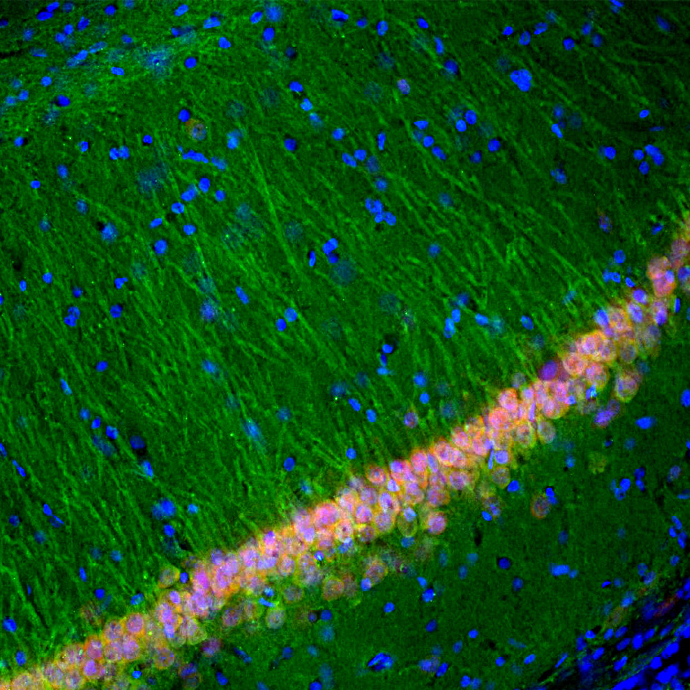
Immunofluorescent analysis of rat hippocampal section stained with mouse mAb to UCHL1, MCA-BH7, dilution 1:5,000 in green and costained with rabbit pAb to FOX/NeuN, RPCA-FOX3, dilution 1:2,000, in red. The blue is Hoechst staining of nuclear DNA. Following transcardial perfusion of rat with 4% paraformaldehyde, brain was post fixed for 24 hours, cut to 45 μM, and free-floating sections were stained with above antibodies. The UCHL1 antibody stains the cell body and dendrites of hippocampal neurons, while the FOX3 antibody labels nuclei of the neuronal cells.
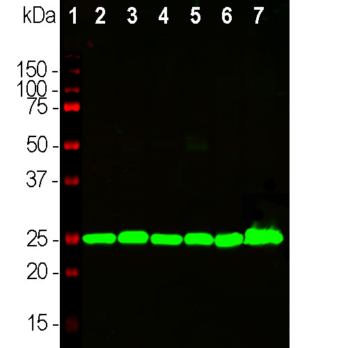
Western blot analysis of tissue lysates using mouse mAb to UCHL1, MCA-BH7, dilution 1:10,000 in green: [1] protein standard (red), [2] rat brain, [3] rat spinal cord, [4] mouse brain, [5] mouse spinal cord, [6] pig brain, [7] pig spinal cord. The single band at 24kDa corresponds to the UCHL1 protein.
Mouse Monoclonal Antibody to UCHL1
Cat# MCA-BH7
Price range: $120.00 through $800.00
Ubiquitin C-terminal hydrolase 1 (UCHL1) is an extremely abundant protein of brain, where it is localized only in neurons. It was originally named PGP9.5 and discovered as a major protein spot on 2D gels of brain extracts which was absent on similar gels of other tissues (1). Later it was found that the PGP9.5 protein was an enzyme which could cleave ubiquitin monomers from ubiquitin conjugates and polyubiquitin chains, resulting in recycling of ubiquitin monomers and the renaming of PGP9.5 to UCHL1 to reflect this enzymatic activity (2). UCHL1 is an essential enzyme and defects in UCHL1 protein expression are involved in Parkinson’s disease (PD) and other more serious disease states (3-6). Genetic studies defined defects in the PARK5 gene as causative of PD in a German family, the PARK5 gene encoding UCHL1 (7). In addition UCHL1 may be released into cerebrospinal fluid (CSF) and blood following CNS damage and disease resulting in neuronal loss. As a result detection of this protein may give information about CNS compromise and recovery (8,9).
The MCA-BH7 antibody was made against full length recombinant human UCHL1 expressed in and purified from E. coli and can be used to identify neurons and their processes in culture or in sections. The immunogen used to generate this antibody is available from EnCor, PROT-r-UCHL1. The antibody works well for western blotting and for IF, ICC and IHC (for IHC see data under “Additional Info” tab). The epitope is centered on the peptide WRFVD, amino acids 26-30 of the human sequence, a region of β structure. Considerable interest has been focused on the detection of UCHL1 in the blood and CSF of patients with traumatic injuries to the brain or spinal cord. This antibody has been widely used as both a capture and a detection reagent in ELISA type assays for measuring UCHL1 levels in blood and CSF samples. In addition EnCor supplies a rabbit polyclonal antibody to UCHL1, RPCA-UCHL1, and also a chicken polyclonal CPCA-UCHL1. We also supply an ELISA kit for the detection of UCHL1 in blood, CSF and other biological fluids, ELISA-UCHL1. Mouse select image at left for larger view.

Chromogenic immunostaining of a 4% PFA fixed paraffin embedded human brain cortex section with mouse mAb to UCHL1, MCA-BH7, dilution 1:10,000, detected in DAB (brown) following the ImmPress method with citra buffer retrieval. Hematoxylin (blue) was used as the counterstain. UCHL1 is extremely abundant in the cell bodies and dendrites in a majority of neuronal cells. This antibody performs well in testing with both 4% PFA and standard NBF fixed human, mouse and rat tissues. Mouse select image for larger view.

Immunofluorescent analysis of cortical neuron-glial culture from E20 rat stained with mouse mAb to UCHL1, MCA-BH7, dilution 1:5,000 in green, and costained with chicken pAb to GFAP, CPCA-GFAP, dilution 1:5,000 in red. The blue is DAPI staining of nuclear DNA. The MCA-BH7 antibody stains cell bodies and dendrites of neurons, while the GFAP antibody labels astrocytes. Mouse select image for larger view.
1. Doran JF, Jackson P, Kynoch PA, Thompson RJ. Isolation of PGP 9.5, a new human neurone-specific protein detected by high-resolution two-dimensional electrophoresis J Neurochem. 40:1542-7 (1983).
2. Wilkinson KD, et al. The neuron-specific protein PGP 9.5 is a ubiquitin carboxyl-terminal hydrolase. Science 246:670-3 (1989).
3. Kurihara LJ, Kikuchi T, Wada K, Tilghman SM. Loss of Uch-L1 and Uch-L3 leads to neurodegeneration, posterior paralysis and dysphagia. Hum. Mol. Genet. 10:1963-70 (2001).
4. Maraganore DM, et al. UCHL1 is a Parkinson’s disease susceptibility gene. Ann Neurol. 55:512-21 (2004).
5. Bilguvar K, et al. Recessive loss of function of the neuronal ubiquitin hydrolase UCHL1 leads to early-onset progressive neurodegeneration. PNAS 110:3489-94 (2013).
6. Liu Y, et al. The UCH-L1 gene encodes two opposing enzymatic activities that affect alpha-synuclein degradation and Parkinson’s disease susceptibility. Cell 111:209-18 (2002).
7. Leroy E, et al. The ubiquitin pathway in Parkinson’s disease. Nature 395:451-2 (1998).
8. Day IN, Thompson RJ. UCHL1 (PGP 9.5): Neuronal biomarker and ubiquitin system protein. Prog. Neurobiol. 90:327-62 (2009).
9. Mondello S, et al. Clinical utility of serum levels of ubiquitin C-terminal hydrolase as a biomarker for severe traumatic brain injury. Neurosurgery 70:666-75 (2012).
Related products


Recently Viewed Products
-
 Mouse Monoclonal Antibody to SARS-CoV2 S-Protein ACE2 Binding Domain
Cat# MCA-5G8 Price range: $120.00 through $800.00 -
 Mouse Monoclonal Antibody to Calretinin
Cat# MCA-6A9 Price range: $120.00 through $800.00 -
 Chicken Polyclonal Antibody to NSE
Cat# CPCA-NSE Price range: $120.00 through $800.00

