| Name: | Mouse monoclonal antibody to rhodopsin |
| Immunogen: | Purified bovine rhodopsin |
| HGNC Name: | RHO |
| UniProt: | P08100 |
| Molecular Weight: | 35kDa |
| Host: | Mouse |
| Isotype: | IgG1 |
| Species Cross-Reactivity: | Human, rat, mouse, cow, pig, horse |
| RRID: | AB_2572379 |
| Format: | Purified antibody at 1mg/mL in 50% PBS, 50% glycerol plus 5mM NaN3 |
| Applications: | WB, IF/ICC, IHC |
| Recommended Dilutions: | WB:1:5,000, IF/ICC and IHC: 1:1,000. |
| Storage: | Store at 4°C for short term, for longer term at -20°C. Avoid freeze / thaw cycles. |
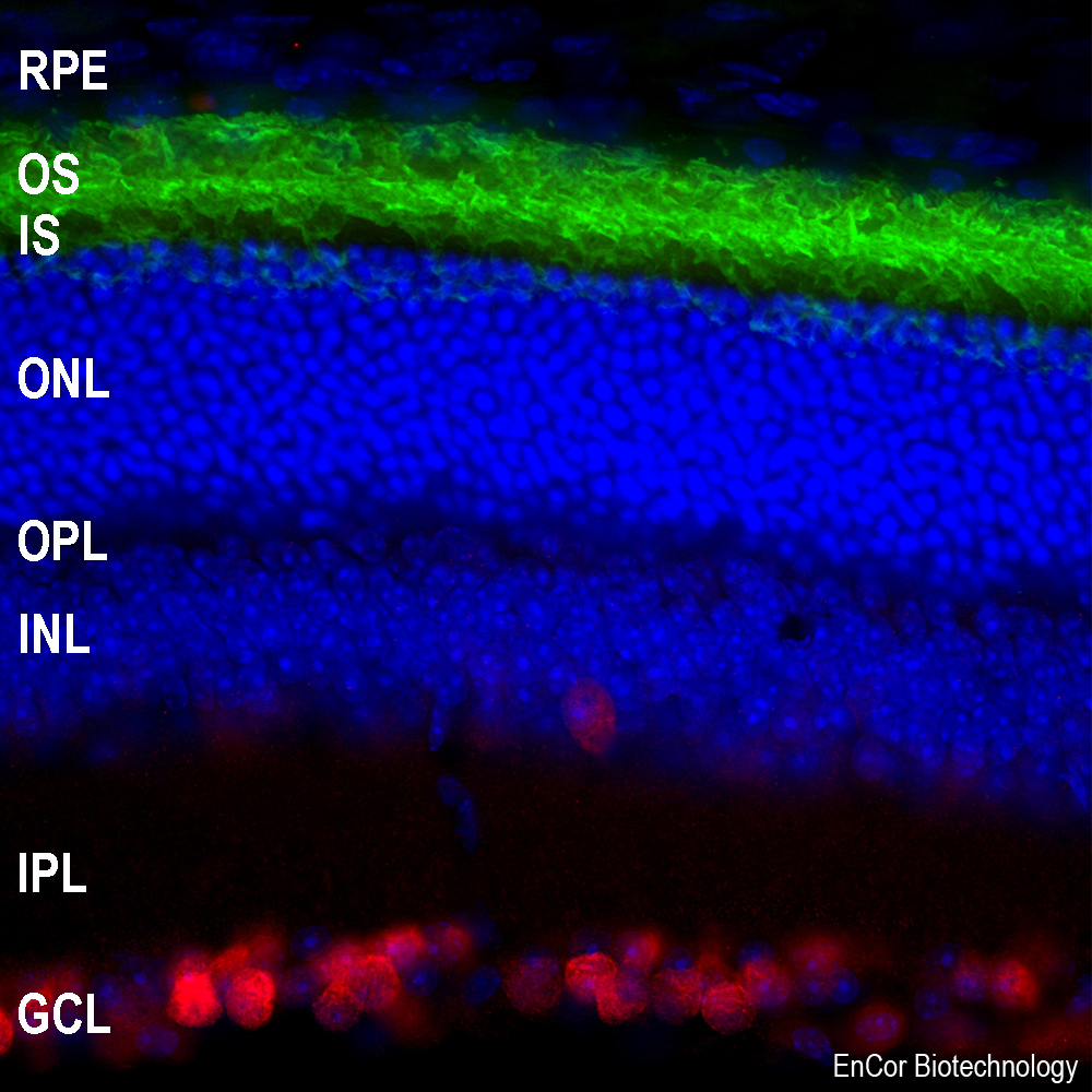
Immunofluorescent analysis of mouse retina section stained with mouse mAb to rhodopsin, MCA-B630, dilution 1:2,000, in green, and costained with rabbit pAb to Fox3/NeuN, RPCA-FOX3, dilution 1:5,000 in red. The blue is Hoechst staining of nuclear DNA. Rhodopsin antibody reveals the rod cell membranes located in photoreceptor outer segments (OS) layer of the retina. The Fox3/NeuN antibody selectively stains the nuclei and cytoplasm of neuronal cells in the ganglion cell layer (GCL), but does not stain most neurons in the layers between.
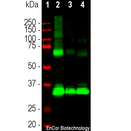
Western blot analysis of retina lysates from different species using mouse mAb to rhodopsin, MCA-B630, dilution 1:5,000 in green: [1] protein standard (red), [2] rat [3] mouse and [4] cow retina lysates. The strong band at 35kDa corresponds to rhodopsin protein. Bands at about 70kDa and 140kDa are presumably aggregated forms of rhodopsin.
Mouse Monoclonal Antibody to Rhodopsin
Cat# MCA-B630
Price range: $120.00 through $800.00
Rhodopsin is the protein in the mammalian retina responsible for the light sensitivity of rod cells which are in turn responsible for vision in low light levels (1-4). Somewhat surprisingly, the rhodopsin protein turned out to be a typical member of the seven transmembrane G protein-coupled receptor (GPCR) superfamily. Whereas other GPCRs initiate signaling on binding a specific ligand, rhodopsin exists with a ligand already bound, specifically the vitamin A related substance retinal. The light causes a conformational change in the receptor bound retinal, which in turn causes a conformational change to the rhodopsin molecule. This change in rhodopsin conformation then results in altered G protein signaling in the rod cell mediated by an inhibitory G protein and ultimately, through several synaptic connections, to low light vision.
The MCA-B630 monoclonal antibody was made in mice against purified rhodopsin from bovine retina (5). The resulting hybridomas were screened first by ELISA on purified bovine rhodopsin. Positive hybridoma were then re-screened on synthetic peptides based on the bovine rhodopsin, so that peptide binding antibodies were epitope mapped, with MCA-B630 binding to a peptide corresponding to the N-terminal 32 amino acids. The antibody works well for western blotting and for IF, ICC and IHC (for IHC see data under “Additional Info” tab). Another mouse monoclonal antibody which has similar properties to MCA-B630, MCA-A531, is also available. Currently MCA-B630 has been more widely used in peer-reviewed studies, although in other respects the two antibodies are comparable. MCA-B630 can be used to study rhodopsin expression both in cell culture, sections and in western blots. Mouse select image at left for larger view.

Chromogenic immunostaining of a 4% PFA fixed paraffin embedded rat retina section with mouse mAb to rhodopsin, MCA-B630, dilution 1:2,000, detected with DAB (brown) using the Vector Labs ImmPRESS method and reagents with citra buffer retrieval. Hematoxylin (blue) was used as the counterstain. In this image, MCA-B630 strongly labels the rod cells within the photo receptor layer of the retina. This antibody has not been tested in NBF fixed material.

High magnification confocal image of pig retinal section stained with MCA-B630 in green. Rhodopsin is most abundant in the rod outer segments (ROS) of retina, clearly localized in rod cell membranes. The rod inner segments (RIS) and rod nuclei in the outer nuclear layer (ONL) are also seen in this image. The blue is Hoechst staining of nuclear DNA.
1. Molday RS. Photoreceptor membrane proteins, phototransduction, and retinal degenerative disease. The Frienwald lecture. Invest Ophthalmol Vis Sci. 39:2491-513 (1998).
2. Yau, KW. Phototransduction Mechanism in Retinal Rods and Cones. The Frienwald lecture. Invest Ophthalmol Vis Sci. 35:9-32 (1994).
3. Wilden U, Hall SW, Kühn H. Phosphodiesterase activation by photoexcited rhodopsin is quenched when rhodopsin is phosphorylated and binds the intrinsic 48-kDa protein of rod outer segments. Proc Natl Acad Sci USA 83:1174-8 (1986).
4. Smith WC, et al. Identification of regions of arrestin that bind to rhodopsin. Biochemistry Mar 38:2752-61 (1999).
5. Adamus G, et al. Use of peptides to select for anti-rhodopsin antibodies with desired amino acid sequence specificities. Pept. Res. 1:42-7 (1988).
This antibody has been widely used in peer-reviewed publications which can be found by searching Google Scholar for “Rhodopsin AND antibody AND B630” or, if you are viewing this online, simply by selecting this link.


