| Name: | Mouse monoclonal antibody to Calreticulin |
| Immunogen: | Synthetic peptides VESGSLEDDWDFLPPKKI corresponding to amino acids 191-208 of human calreticulin, including the LC3 interacting region or LIR. |
| HGNC Name: | CALR |
| UniProt: | P27797 |
| Molecular Weight: | 48kDa |
| Host: | Mouse |
| Isotype: | IgG1 |
| Species Cross-Reactivity: | Human, Rat, Mouse |
| RRID: | AB_2572240 |
| Format: | Purified antibody at 1mg/mL in 50% PBS, 50% glycerol plus 5mM NaN3 |
| Applications: | WB, IF/ICC, IHC |
| Recommended Dilutions: | WB: 1:1,000-1:2,000 IF/ICC 1:1,000 IHC: 1:500. |
| Storage: | Store at 4°C for short term, for longer term at -20°C |
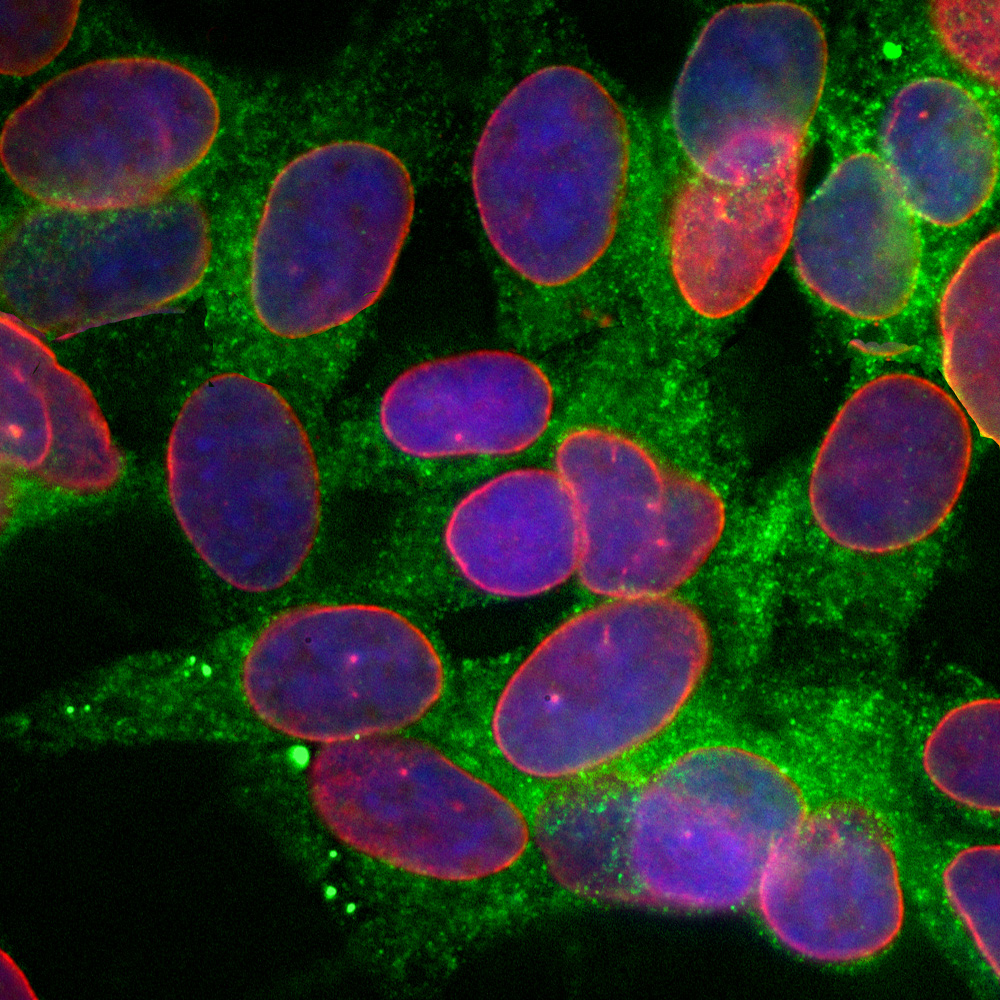
Immunofluorescent analysis of of SH-SY5Y cells stained with mouse mAb to calreticulin, MCA-6C6, dilution 1:500 in green and costained with chicken pAb to lamin A/C, CPCA-LaminAC, dilution 1:2,000 in red. The blue is DAPI staining of nuclear DNA. The MCA-6C6 antibody reveals granular staining of cytoplasm, while the lamin A/C antibody stains the nuclear lamina and membrane.
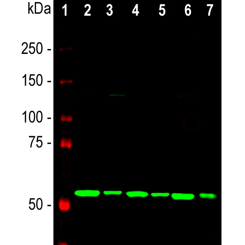
Western blot analysis of lysates of different cell lines using mouse mAb to calreticulin, MCA-6C6, dilution 1:2,000: [1] protein standard, [2] NIH-3T3, [3] HEK293, [4] HeLa, [5] SH-SY5Y, [6] C6, [7] COS-1 cells. A strong single band at about 50kDa corresponds to the calreticulin protein.
Mouse Monoclonal Antibody to Calreticulin
Cat# MCA-6C6
Price range: $120.00 through $800.00
Calreticulin was first identified as a calcium binding protein found in rabbit skeletal muscle. It appears to be a multifunctional protein found predominantly in endoplasmic reticulum (ER) and is ubiquitously expressed in a wide range of species (1). The protein has a conserved globular ~170 amino acid lectin-like N-terminal domain, a central proline rich region and a C-terminal low affinity but high capacity acidic Calcium binding region. There is a four-amino acid ER retention sequence (KDEL) at the extreme C-terminus accounting for ER localization. Gene knock out experiments show that the absence of the calreticulin gene is embryonically lethal (2). One of the major functions of calreticulin appears to be buffering Calcium levels in the ER which also affects intracellular calcium signaling (3). Calreticulin also functions as an autophagy receptor and as a molecular chaperone for glycoproteins (4). Calreticulin may also be found on the cell surface where it may mediate phagocytosis of apoptotic or dying tumor cells by triggering immune responses and allowing the immune system to remove tumor cells. This suggests that targeting cell surface calreticulin may be a novel anti-tumor therapy (5-8). The protein includes a functional “LC3 interacting region” or LIR motif (9). Proteins with LIR motifs bind ATG8 family members which in mammals are the LC3 and GABARAP proteins and calreticulin has been shown to bind binds LC3 (10). This binding is one of the essential steps in the process of autophagy, so LIR motif proteins generally function to target other proteins which bind to them for degradation.
The MCA-6C6 antibody was raised against the peptide VESGSLEDDWDFLPPKKI, corresponding to amino acids 191-208 of the human calreticulin protein. This peptide includes the LIR motif of the molecule, centered on the peptide WxxL, so this antibody may block binding of other molecules to this site (9,11). The antibody works well for western blotting and for IF, ICC and IHC (for IHC see data under “Additional Info” tab). Mouse select image above left for larger view.

Chromogenic immunostaining of a formalin fixed paraffin embedded human brain cortex section with mouse mAb to calreticulin, MCA-6C6, dilution 1:500, detected with DAB (brown) using the Vector Labs ImmPRESS method and reagents with citra buffer retrieval. Hematoxylin (blue) was used as the counterstain. In this image, MCA-6C6 intensely labels the cytoplasm of pyramidal neurons. This antibody performs well in testing with both 4% PFA and standard NBF fixed tissues from human and rat. Mouse select image for larger view.
1. Ostwald TJ, MacLennan DH. Isolation of a high affinity calcium-binding protein from sarcoplasmic reticulum. J. Biol. Chem. 249:974-9 (1974).
2. Mesaeli N, et al. Calreticulin is essential for cardiac development. J. Cell Biol. 144:857–68 (1999).
3. Michalak M, et al. Calreticulin, a multi-process calcium-buffering chaperone of the endoplasmic reticulum. Biochem. J. 417:651–66 (2009).
4. Bedard K, Szabo E, Michalak M, Opas M. Cellular functions of endoplasmic reticulum chaperones calreticulin, calnexin, and ERp57. Int. Rev. Cytol. 245:91–121 (2005).
5. Obeid M, et al. Calreticulin exposure dictates the immunogenicity of cancer cell death. Nat. Med. 13:54–61 (2007).
6. Tesniere A, et al. Immunogenic cancer cell death: a key-lock paradigm. Curr. Opin. Immunol. 20:504–11 (2008).
7. Gardai SJ, et al. Cell-surface calreticulin initiates clearance of viable or apoptotic cells through trans-activation of LRP on the phagocyte. Cell 123:321–34 (2005).
8. Fucikova J, et al. Relevance of the chaperone-like protein calreticulin for the biological behavior and clinical outcome of cancer. Immunol. Lett. 193:25-34 (2018).
9. Birgisdottir AB, Lamark T, Johansen T. The LIR motif – crucial for selective autophagy. J. Cell Sci. 126:3237–47 (2013).
10. Yang Y, et al. The ER-localized Ca 2+-binding protein calreticulin couples ER stress to autophagy by associating with microtubule-associated protein 1A/1B light chain 3. J. Biol. Chem. 294:772-782 (2019).
11. Slobodkin MR, Elazar Z. The Atg8 family: multifunctional ubiquitin-like key regulators of autophagy. Essays Biochem. 55:51-64 (2013).
Related products


Recently Viewed Products
-
 Mouse Monoclonal Antibody to GAP43
Cat# MCA-5E8 Price range: $120.00 through $800.00 -
 Mouse Monoclonal Antibody to HMGB1
Cat# MCA-1F3 Price range: $120.00 through $800.00 -
 Mouse Monoclonal Antibody to Arrestin1
Cat# MCA-S128 Price range: $120.00 through $800.00

