| Name: | Mouse monoclonal antibody to TDP43 |
| Immunogen: | Full length recombinant human TDP43 expressed in and purified from E. coli. |
| HGNC Name: | TARDBP |
| UniProt: | Q13148 |
| Molecular Weight: | 43kDa |
| Host: | Mouse |
| Isotype: | IgG1 heavy, κ light |
| Species Cross-Reactivity: | Human, rat, mouse, cow, pig, horse |
| RRID: | AB_2572387 |
| Format: | Purified antibody at 1mg/mL in 50% PBS, 50% glycerol plus 5mM NaN3 |
| Applications: | WB, IF/ICC, IHC |
| Recommended Dilutions: | WB: 1:5,000. ICC/IF and IHC 1:1,000 |
| Storage: | Store at 4°C for short term, for longer term at -20°C. |
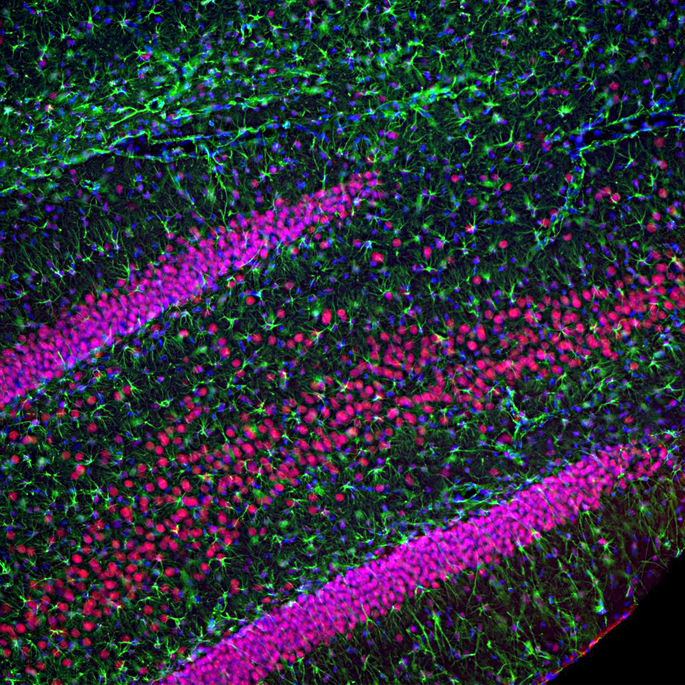
Immunofluorescent analysis of rat hippocampus section stained with mouse mAb to TDP43, MCA-3H8, dilution 1:2,000 in red, and costained with chicken pAb to GFAP, CPCA-GFAP, dilution 1:5,000 in green. The blue is DAPI staining of nuclear DNA. Following transcardial perfusion of rat with 4% paraformaldehyde, brain was post fixed for 24 hours, cut to 45μM, and free-floating sections were stained with above antibodies. The TDP43 protein is concentrated in the nuclei of hippocampal neurons, while the GFAP antibody stains the intermediate filament network of astroglial cells.
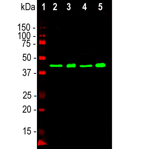
Western blot analysis of whole brain lysates and nuclear extract from whole brain using mouse mAb to TDP43, MCA-3H8, dilution 1:2,000 in green: [1] protein standard (red), [2] rat brain, [3] rat brain nuclear extract, [4] mouse brain, [5] mouse brain nuclear extract. Strong band at the 43kDa mark visible both in whole brain and in the nuclear extract corresponds to TDP43 protein.
Mouse Monoclonal Antibody to TDP43
Cat# MCA-3H8
Price range: $120.00 through $800.00
TDP43 was originally identified as a protein which binds to the “transactivation response” (TAR) sequence found in the long terminal repeat of the HIV-1 virus genome (1). UV cross-linking of HeLa cell extract revealed a 43kDa protein which was cloned and sequenced and shown to contain a two central copies of the ~90 amino acid RNA recognition motif or RRM domain. RRM is an acronym for RNA Recognition Motif, and this domain is found in many proteins which bind single stranded RNA and some which bind single stranded DNA. It also has a single bipartite nuclear localization sequence in the N-terminus region in front of the first RRM domain, and the C-terminal region, beyond the second RRM domain is rich in glycine, asparagine and glutamine. TDP43 is normally expressed in the mammalian CNS but accumulates in nuclear inclusions in the neurons of patients with frontotemporal lobar degeneration (FTLD), amyotrophic lateral sclerosis (ALS), Alzheimer’s disease and other pathologies (2-4). Disease related point mutations in TDP43 are almost always found in the C-terminal region. TDP43 is related in protein sequence to FUS/TLS, EWSR1 and TAF15 and all four proteins have a tendency, especially when mutated, to aggregate into inclusions and are implicated in a variety of neurological disorders (5,6).
The MCA-3H8 antibody was made against full length recombinant human protein and works well on western blots, by IF, ICC and IHC, staining normal neurons and FTLD and ALS related inclusion bodies, see “Additional Info” tab. It has also been shown to identify inclusions seen in Alzheimer’s disease (4). We have shown that MCA-3H8 reacts with TDP43 from human, cow, pig, mouse, rat and other mammals and that the epitope is in the center the peptide HNSNRQLERSGRFGGNPGGF, amino acids 264-283, C-terminal to second RRM domain of the human sequence. This is the region typically in TDP43 inclusions (see here). This peptide is 100% conserved in a wide variety of mammalian species so it is likely that the antibody is effective on many other species also. The KD for this antibody in 1.16 X 10⁻⁹M. The antibody is also an excellent marker of nuclei in cell fractionation experiments. Mouse select image at left for rather view, and on “additional info” tab for more information.

Chromogenic immunostaining of a 4% PFA fixed paraffin embedded rat cerebellum section with mouse mAb to TDP43, MCA-3H8, dilution 1:4,000, detected with DAB (brown) using the Vector Labs ImmPRESS method and reagents with citra buffer retrieval. Hematoxylin (blue) was used as the counterstain. The MCA-3H8 antibody strongly and specifically labels nuclei. This antibody performs well in testing with both 4% PFA and standard NBF fixed tissues.Mouse select image for larger view.

The mouse monoclonal antibody to TDP43, MCA-3H8, is a robust marker of TDP43 positive inclusions in ethanol fixed and paraffin embedded sections, as shown here on a section of human brain from a patient with fronto-temporal dementia (FTLD). Image courtesy of Dr. Benoit Giasson of the Center for Translational Research in Neurodegenerative Disease (CTRND) at the University of Florida. Mouse select image for a larger view.
1. Ou SH. et al. Cloning and characterization of a novel cellular protein, TDP-43, that binds to human immunodeficiency virus type 1 TAR DNA sequence motifs. J Virol. 69:3584-96 (1995).
2. Neumann M. et al. Ubiquitinated TDP-43 in frontotemporal lobar degeneration and amyotrophic lateral sclerosis. Science 314:130-33 (2006).
3. Forman MS, Trojanowski JQ and Lee VM-Y. TDP-43: a novel neurodegenerative proteinopathy. Curr. Opin. Neurobiol. 17:548-55 (2007).
4. Wilson AC, Dugger BN, Dickson DW and Wang DS. TDP-43 in aging and Alzheimer’s disease – a review. Int. J. Clin. Exp. Pathol. 4:147-55 (2011).
5. Da Cruz S, Cleveland DW. Understanding the role of TDP-43 and FUS/TLS in ALS and beyond. Curr. Opin. Neurobiol. 21:904-19 (2011).
6. Couthouis J, et al. A yeast functional screen predicts new candidate ALS disease genes. PNAS 108:20881-90 (2011).
The antibody has also been sold through many OEM partners, and peer-reviewed publications making use of it can be found by searching Google Scholar for “3H8 AND TDP43 AND antibody” or simply by selecting this link.


