| Name: | Mouse Monoclonal to GAP43 MCA-1E3 |
| Immunogen: | C-terminal peptide of rat and mouse GAP43, which is KEDPEADQEHA, with an N-terminal Cys added to allow chemical coupling to KLH carrier protein. |
| HGNC Name: | GAP43 |
| UniProt: | P07936 |
| Molecular Weight: | 38, 43kDa by SDS-PAGE |
| Host: | Mouse |
| Isotype: | IgG1 |
| Species Cross-Reactivity: | Human, rat, mouse |
| RRID: | AB_2572285 |
| Format: | Affinity purified at 1mg/mL in PBS, 50% glycerol, 5mM NaN3 |
| Applications: | WB, IF/ICC, IHC |
| Recommended Dilutions: | WB: 1:10,000. IF/ICC and IHC: 1:1,000. |
| Storage: | For long term storage, leave frozen at -20°C. Avoid freeze / thaw cycles. |
Mixed neuronal cultures stained with MCA-1E3 (green), RPCA-MAP2, a rabbit antibody to microtubule associated protein 2 (MAP2, red) and DNA (blue). The GAP43 antibody stains the plasma membrane of neurons and is particularly concentrated in dendrites. Another image is here.
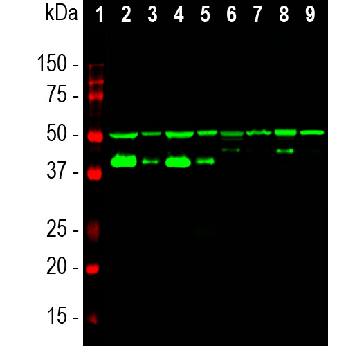
Western blot analysis of different tissue lysates using mouse mAb to GAP43, MCA-1E3, dilution 1:10,000 in green: [1] protein standard (red), [2] rat brain, [3] rat spinal cord, [4] mouse brain, [5] mouse spinal cord, [6] pig hippocampus, [7] pig spinal cord,[8] cow cerebellum, and [9] cow spinal cord. 2 bands at 43 and 50kDa mark correspond to alternative transcripts of GAP43 protein.
Mouse Monoclonal Antibody to GAP43
Cat# MCA-1E3
Price range: $120.00 through $800.00
GAP43 is an abundant protein which is found heavily concentrated in developing neurons, in particular at the growing tips, the growth cones. One group discovered it since it becomes unregulated during the regeneration of the toad optic nerve, and named it “growth associated protein 43”, the 43 referring to the apparent molecular weight on SDS-PAGE gels (1). GAP43 does not run on SDS-PAGE in a fashion which accurately reflects its molecular weight, since the full length molecule human GAP43 is 238 amino acids giving a real molecular weight 24.8kDa. The molecule is very rich in charged and has few large hydrophobic amino acids which causes it to bind SDS poorly. The same GAP43 preparation will also give a different SDS-PAGE molecular weight depending on the percentage acrylamide content of the gel, the protein appearing relatively larger on gels with higher acrylamide concentration. GAP43 proteins from different species also may run at different apparent molecular weights on the same gel. Partly due to these unusual features GAP43 was independently discovered by several different groups and therefore has several alternate names, such as protein F1, pp46, neuromodulin, neural phosphoprotein B-50 and calmodulin-binding protein P-57, the numbers 46, 50 and 57 reflecting the apparent SDS-PAGE molecular weight (2). GAP43 is a major protein kinase C substrate and binds calmodulin avidly, this being mediated by an N-terminal IQ calmodulin binding motif (3). GAP43 may be anchored to the plasma membrane by reversible palmitoylation on two Cys residues close to the N-terminus (4). Knock out of the GAP43 gene in mice is lethal early in postnatal life and is associated with defects in axonal pathfinding (5). GAP43 is one of a large family of “intrinsically disordered proteins” which typically have little defined structure unless they are bound to a more structured partner (6).
The MCA-1E3 antibody was made against the C-terminal peptide of rat GAP43 and binds to GAP43 in rodents and other mammalian species. It binds strongly to growth cones and axonal processes of neurons in cell culture and to synaptic regions in sectioned material. It also works well for western blotting and IHC, see data under the “Additional Info” tab. We also supply rabbit and chicken polyclonal antibodies to GAP43, RPCA-GAP43 and CPCA-GAP43 respectively. Mouse select image at left for larger view.

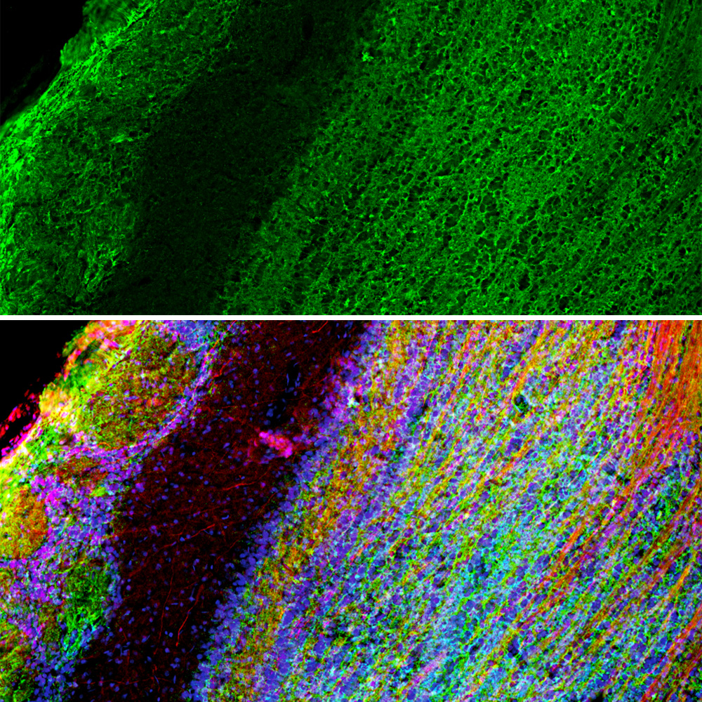
Chromogenic immunostaining of a NBF fixed paraffin embedded human cerebellum section with mouse mAb to GAP43, MCA-1E3, dilution 1:2,000, detected in DAB (brown) using Vector Labs ImmPRESS method and reagents with citra buffer retrieval. Hematoxylin (blue) was used as the counterstain. The GAP43 antibody labels molecular layer cells and neuropil as well as processes in the granular layer. This antibody performs well in testing with both 4% PFA and standard NBF fixed tissues. Mouse select image for larger view.
1. Skene JH, Willard M. Changes in axonally transported proteins during axon regeneration in toad retinal ganglion cells.J. Cell Biol. 89:86-95 (1981).
2. Benowitz LI, Routtenberg A. GAP-43: an intrinsic determinant of neuronal development and plasticity. Trends Neurosci. 20:84-91 (1997).
3. Kosik KS, et al. Human GAP-43: its deduced amino acid sequence and chromosomal localization in mouse and human. Neuron 1:137-32 (1988).
4. Gauthier-Kempera A, et al. Interplay between phosphorylation and palmitoylation mediates plasma membrane targeting and sorting of GAP43. Mol Biol Cell. 25:3284-99 (2014).
5. Strittmatter SM, et al. Neuronal pathfinding is abnormal in mice lacking the neuronal growth cone protein GAP-43. Cell 80:445-52 (1995).
6. Wright PE. Dyson HJ. Intrinsically disordered proteins in cellular signalling and regulation. Nat. Rev. Mol. Cell Biol. 16:18-29 (2015).
Related products


Recently Viewed Products
-
 Rabbit Polyclonal Antibody to Ankyrin 3/Ankyrin G
RPCA-ANK3 Price range: $150.00 through $1,000.00

