| Name: | Mouse Monoclonal Antibody to Adenylate Cyclase III (ACIII) |
| Immunogen: | C-terminal peptide of rat ACIII, PAAFPNGSSVTLPHQVVDNP with a Cys added to the N-terminus to allow coupling to KLH. |
| HGNC Name: | ADCY3 |
| UniProt: | P21932 |
| Molecular Weight: | ~120kDa and above |
| Host: | Mouse |
| Isotype: | IgG1 |
| Species Cross-Reactivity: | Human, Rat, Mouse |
| RRID: | AB_2744501 |
| Format: | Purified antibody at 1mg/mL in 50% PBS, 50% glycerol plus 5mM NaN3 |
| Applications: | WB, IF/ICC, IHC |
| Recommended Dilutions: | WB: 1:1,000-1:2,000. IF/ICC and IHC: 1:1,000 |
| Storage: | Store at 4°C for short term, for longer term at -20°C |
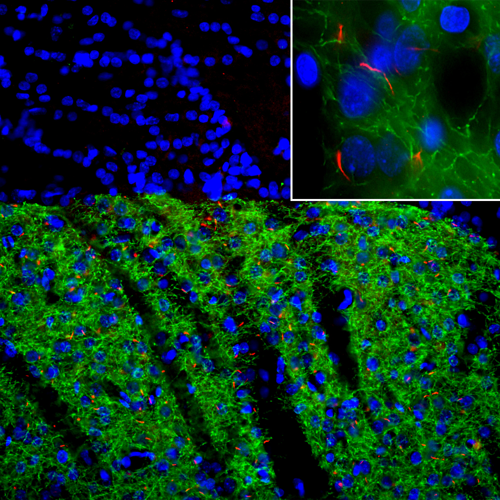
Immunofluoresence of caudate/putamen region of rat brain stained with mouse mAb to ACIII, MCA-1A12, in red, and chicken antibody to tyrosine hydroxylase, in green. The Blue is Hoechst stain revealing nuclei. The ACIII antibody reveals neuronal cilia while the tyrosine hydroxylase antibody reveals the axons of chatecholaminergic neurons.
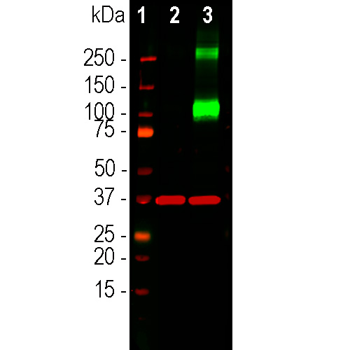
Western blot analysis of HEK293 cell lysates using mouse mAb to ACIII, MCA-1A12, dilution 1:1,000, in green: [1] protein standard, [7] non-transfected HEK293 cells, and [8] HEK293 cells transfected with a vector encoding Myc-DDK tagged fill length human adenylate cyclase III (ACIII). The strong band at about 130kDa in transfected cells demonstrates overexpression of the human ACIII protein, and bands over 250kDa presumably correspond to either heavily glycosolated form of ACIII or multimeric forms of the protein. The same blot was simultaneously probed with rabbit pAb to GAPDH, RPCA-GAPDH, dilution 1:20,000 in red, which reveals the single band at ~37kDa seen in both transfected and non-transfected cells.
Mouse Monoclonal Antibody to ACIII
Cat# MCA-1A12
Price range: $120.00 through $800.00
Trimeric G-proteins are a large and variable family of membrane receptors. On binding their specific ligand they activate specific members of the family of trimeric G-proteins which in turn activate other signalling enzymes. Adenylate cyclases are one of these downstream enzyme families which are activated by the GTP bound GαS subunits of trimeric G-proteins. Adenylate cyclases are responsible for the production of the important “second messenger” signaling molecule cyclic-AMP which in turn activates the cAMP dependent protein kinase. This kinase when activated phosphorylates numerous substrate molecules on serine or threonine residues and so alters their activity. There are several different adenylate cyclase genes and protein products with each have distinctly different distribution patterns in cells and tissues. The type III adenylate cyclase enzyme is specifically localized in the membranes surrounding neuronal cilia, and is activated by specific G-protein coupled receptors also located in cilia (1-5). Neuronal cilia express a variety of other receptors types and mediators of other signaling pathways and appear to function as a unique and complex neuronal sensory structure (1-5). For examples, the somatostatin 3 receptor, neuropeptide Y 2 receptor and melanin concentrating hormone receptor 1 are localized in neuronal cilia and the sonic hedgehog and Wnt signalling pathway act on neurons primarily through neuronal cilia (6). This antibody is an excellent marker of neuronal cilia in the brain and in cells in tissue culture and works in the same way as our rabbit polyclonal made against the same peptide (7). The antibody was recently utilized in a very high profile publication in the journal Cell (8).
The MCA-1A12 antibody was made against the extreme C-terminal peptide of rat ACIII, PAAFPNGSSVTLPHQVVDNP, amino acids 1125-1144 of the Genbank entry NP_570135.2. A cysteine residue was added to the N-terminus to allow coupling to MBS-activated keyhole limpet hemocyanin. The antibody works on mouse cells which express the same peptide and also on human cells, presumably because the corresponding peptide in the human AC3 sequence is the closely related peptide LATFPNGPSVTLPHQVVDNS. The antibody works well to identify neuronal cilia on both human and rodent cells by IF and ICC but is not recommended for IHC. We have also generated rabbit and chicken polyclonal antibodies to the same ACIII peptide, RPCA-ACIII and CPCA-ACIII. Mouse select image at left for larger view.
1. Fuchs JL, Schwark HD. Neuronal primary cilia: a review. Cell Biol. Int. 28:111-8 (2004).
2. Louvi A and Grove EA. Cilia in the CNS: the quiet organelle claims center stage. Neuron 69:1046-60 (2011).
3. Singla V, Reiter JF. The primary cilium as the cell’s antenna: signaling at a sensory organelle. Science 313:629-33 (2006).
4. Green JA, Mykytyn K. Neuronal Primary Cilia: An Underappreciated Signaling and Sensory Organelle in the Brain. Neuropsychopharm. 39:244–5 (2014).
5. May-Simera HL, Kelley MW. Cilia, Wnt signaling, and the cytoskeleton. Cilia 2;1:7 (2012).
6. Guemez-Gamboa A, et al. Primary cilia in the developing and mature brain. Neuron 82:511-21 (2014).
7. Guadiana SM, et al. Arborization of Dendrites by developing neocortical neurons is dependent on primary cilia and Type 3 adenylyl cyclase. J. Neurosci. 33:2626-38 (2013).
8. Sheu S-H et al. A serotonergic axon-cilium synapse drives nuclear signaling to alter chromatin accessibility. Cell 185:3390-3407 (2022).
Related products


Recently Viewed Products
-
 Chicken Polyclonal Antibody to GFAP
Cat# CPCA-GFAP Price range: $120.00 through $800.00

