| Name: | Chicken polyclonal antibody to neurofilament NF-M |
| Immunogen: | Recombinant construct containing the C-terminus of the human sequence (amino acids 708-877) expressed in and purified from E. coli. |
| HGNC Name: | NEFM |
| UniProt: | P07197 |
| Molecular Weight: | 145-160kDa by SDS-PAGE |
| Host: | Chicken |
| Isotype: | |
| Species Cross-Reactivity: | Human, rat, mouse, cow, pig, horse, chicken |
| RRID: | AB_2572367 |
| Format: | Concentrated IgY preparation in PBS plus 0.02% NaN3 |
| Applications: | WB, IF/ICC, IHC |
| Recommended Dilutions: | WB: 1:2,000-5,000, IF/ICC & IHC: 1:2,000 |
| Storage: | Store at 4°C for short term, for longer term store at -20°C |
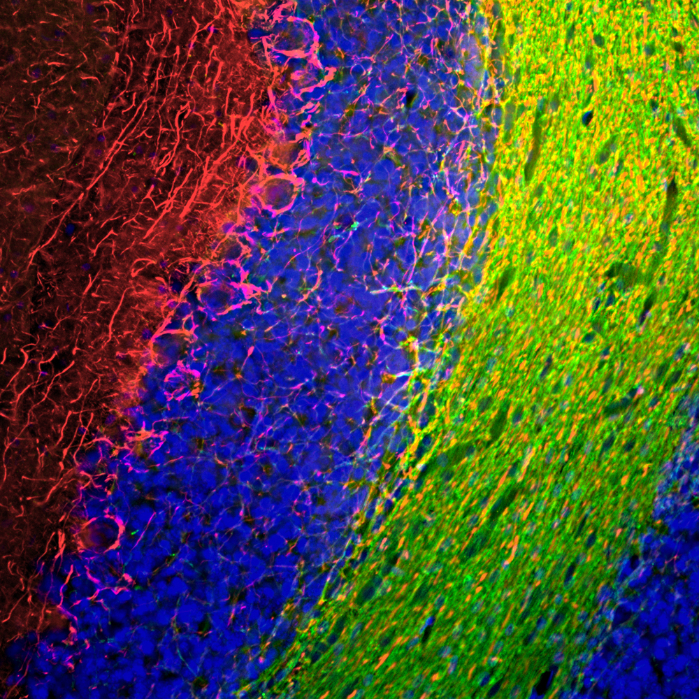
Immunofluorescent analysis of rat cerebellum section stained with chicken pAb to NF-M, CPCA-NF-M, dilution 1:1,000 in red, and costained with mouse mAb to CNP, MCA-1H10, dilution 1:500 in green. The blue is DAPI staining of nuclear DNA. Following transcardial perfusion of rat with 4% paraformaldehyde, brain was post fixed for 24 hours, cut to 45μM, and free-floating sections were stained with the above antibodies. The NF-M antibody labels the network of axons of basket neurons and other neurons. The CNP antibody stains oligodendrocytes, cells that create myelin sheaths around axons.
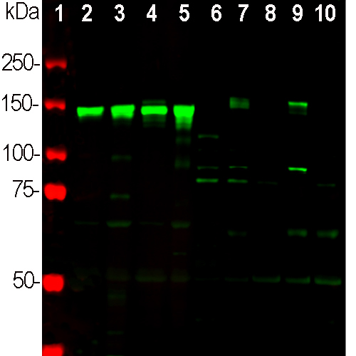
Western blot analysis of different neuronal tissue and cell lysates using chicken pAb to NF-M, CPCA-NF-M, dilution 1:2,000 in green: [1] protein standard (red), [2] rat brain [3] rat spinal cord, [4] mouse brain, [5] mouse spinal cord, [6] NIH/3T3 cells, [7] HEK293, [8] HeLa, [9] SH-SY5Y, and [10] C6 cells. Strong band at 145kDa corresponds to rodent NF-M, and about 160kDa band corresponds to human NF-M protein, visible in SHSY-5Y and HEK293 cells which have neuronal properties. NF-M is not expressed in HeLa and other cell lines tested.
Chicken Polyclonal Antibody to Neurofilament NF-M
Cat# CPCA-NF-M
Price range: $120.00 through $800.00
Neurofilaments are the 10nm or intermediate filament proteins found specifically in neurons, and are composed predominantly of three major proteins called NF-L, NF-M and NF-H. NF-M is the neurofilament middle or medium molecular weight polypeptide and runs on SDS-PAGE gels at 145-160kDa, with some species variability, though the real molecular weight is ~105kDa. The major function of neurofilaments is likely to control the diameter of large axons (1). Antibodies to NF-M such as CPCA-NF-M are useful for identifying neuronal cells and their processes in tissue sections and in cell culture. NF-M antibodies can also be useful to visualize neurofilament rich accumulations seen in many neurological diseases, such as Amyotrophic Lateral Sclerosis (a.k.a. Lou Gehrig’s disease) and Alzheimer’s disease (2-4). Much recent evidence has suggested that the detection of NF-L and NF-H in blood and CSF might be a useful prognostic or diagnostic biomarkers of neuronal damage and degeneration associated with a variety of CNS pathologies (5,6). The potential utility of NF-M in this fashion has not to date been examined.
The CPCA-NF-M antibody was made against a recombinant fusion protein of E. coli TrpE fused to the C-terminus of rat NF-M, amino acids 677-845 (7). This region is very highly conserved in protein sequence across species boundaries and contains some interesting peptide repeats of currently unknown function (8). The CPCA-NF-M antibody is very similar in properties to a rabbit polyclonal the production and characterization of which were described in reference 7. Also available from EnCor is a rabbit polyclonal and a widely used mouse monoclonal antibody to the same immunogen RPCA-NF-M, and MCA-3H11. All three antibodies works on a variety of species and are clean and specific on western blots, cell and tissue staining. Mouse select image at left for larger view.

Chromogenic Immunostaining of a formalin fixed paraffin embedded human cerebellum with chicken pAb to NF-M, CPCA-NF-M, dilution 1:2,000, detected in DAB (brown) following the ABC method. Hematoxylin (blue) was used as the counterstain. The NF-M antibody detects perikarya and dendrites of neuronal Purkinje cells, and is strongly expressed in the axons of basket and other kinds of neuron. Mouse select image for larger view.
1. Hoffman et al. Neurofilament gene expression:a major determinant of axonal caliber. PNAS 84:3472-6 (1987).
2. Perrot R, et al. Review of the Multiple Aspects of Neurofilament Functions, and their Possible Contribution to Neurodegeneration. Mol. Neurobiol. 38:27-65 (2008).
3. Lépinoux-Chambaud C. Eyer J. Review on intermediate filaments of the nervous system and their pathological alterations. Histochem. Cell Biol. 140:13-22 (2013).
4. Liu Q. et al. Neurofilamentopathy in Neurodegenerative Diseases. Open Neurol. J. 5:58–62 (2011).
5. Bacioglu M, et al. Neurofilament light chain in blood and CSF as marker of disease progression in mouse models and in neurodegenerative diseases. Neuron 91:56-66 (2016).
6. Shaw G. The use and potential of pNF-H as a general blood biomarker of axonal loss: an immediate application for CNS injury. in Brain Neurotrauma: Molecular, Neuropsychological, and Rehabilitation Aspects. CRC Press/Taylor & Francis Chapter 21 (2015).
7. Harris J, Ayyub C. and Shaw G. A molecular dissection of the carboxyterminal tails of the major neurofilament subunits NF-M and NF-H. J. Neurosci. Res. 30:47-62 (1991).
8. Shaw G. Identification of previously unrecognized sequence motifs at the extreme carboxyterminus of the neurofilament subunit NF-M. BBRC 14;162:294-9 (1989).
Related products


Recently Viewed Products
-
 Mouse Monoclonal Antibody to Calretinin
Cat# MCA-6A9 Price range: $120.00 through $800.00
