| Name: | Chicken Polyclonal Antibody to mCherry |
| Immunogen: | Full length recombinant protein expressed in and purified from E. coli. |
| HGNC Name: | N.A. |
| UniProt: | D1MPT3 |
| Molecular Weight: | ~28kDa |
| Host: | Chicken |
| Isotype: | |
| Species Cross-Reactivity: | Not Applicable |
| RRID: | AB_2572308 |
| Format: | Concentrated IgY preparation in PBS plus 0.02% NaN3 |
| Applications: | WB, IF/ICC, IHC |
| Recommended Dilutions: | WB: 1:2,000-5,000 IF/IHC: 1:1,000. |
| Storage: | Stable at 4°C |
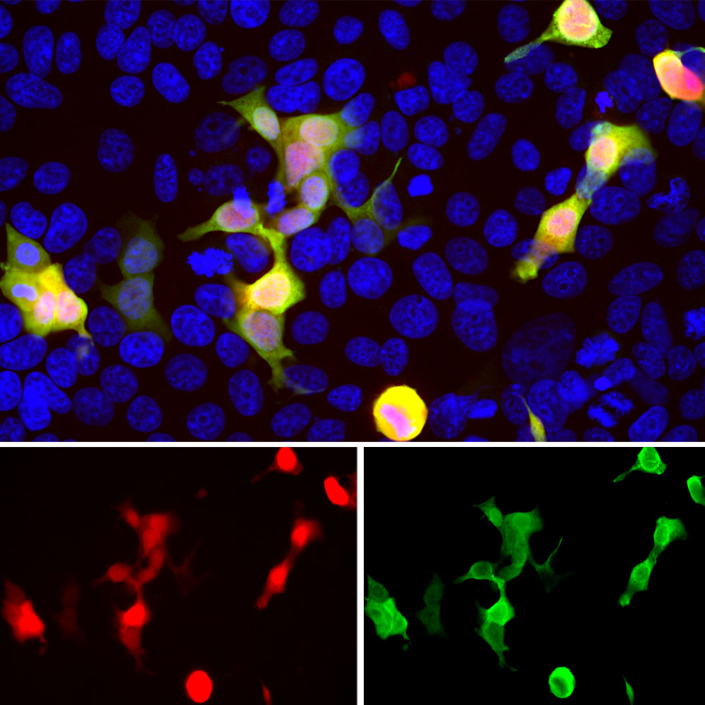
HEK293 cells transfected with a mCherry, stained with CPCA-mCherry antibody and viewed in a confocal microscope. Most HEK293 cells are not transfected so only the nucleus of these cells can be visualized with a blue DNA stain. Cells which are transfected with mCherry are bright red, and staining with CPCA-mCherry is shown in Green. The green antibody staining is only seen cells which express mCherry, as expected, and the superimposition of the green and red results in an orange signal. Interestingly stronger mCherry staining is seen in the nucleus, possibly due to degradation of some mCherry molecules releasing the low molecular weight mCherry fluorochrome.
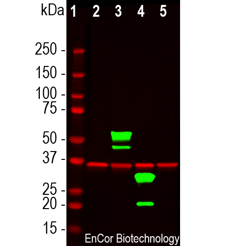
Western blot analysis of HEK293 cell lysates using chicken pAb to mCherry, CPCA-mCherry, dilution 1:2,000, in green, and rabbit pAb to GAPDH, RPCA-GAPDH, dilution 1:5,000, in red: [1] protein molecular weight standard, [2] untransfected HEK293 control cells, [3] HEK293 cells transfected with pCI-Neo-mod vector expressing two tdTomato protein domains, [4] HEK293 cells transfected with pCI-Neo-mod vector expressing one mCherry-HA protein domain, and [5] HEK293 cells transfected with pCI-Neo-mod vector expressing one GFP domain. The CPCA-mCherry antibody recognizes tdTomato and mCherry proteins revealing major bands at about 60kDa and 30kDa, but does not recognize GFP. The red band at 37kDa corresponds to GAPDH protein here used as a loading control.
Chicken Polyclonal Antibody to mCherry
Cat# CPCA-mCherry
Price range: $120.00 through $800.00
mCherry is derived from proteins originally isolated from Cnidarians (jelly fish, sea anemones and corals), and is used as a fluorescent tracer in transfection and transgenic experiments. The prototype for these fluorescent proteins is green fluorescent protein (GFP), which is a ~27kDa protein isolated originally from the jellyfish Aequoria victoria. GFP was the basis of the 2008 Nobel prize in chemistry, specifically “for the discovery and development of the green fluorescent protein, GFP”. GFP and also mCherry fluoresce on contact with molecular oxygen, requiring no other cofactors, and so can be expressed in fluorescent form in essentially any prokaryotic or eukaryotic cell. The mCherry protein is derived from DsRed, a red fluorescent protein from so-called disc corals of the genus Discosoma. DsRed is similar in size and properties to GFP, but, obviously, produces a red rather than a green fluorochrome. The original DsRed was engineered extensively in the Tsien lab to prevent it from forming tetramers and dimers and to modify and improve the spectral properties. Several further cycles of mutation, directed modification and evolutionary selection produced mCherry, which has an excitation maximum at 587nm and emission maximum at 610nm.The mCherry antibody can used to verify the correct size of mCherry fusions by western blotting in extracts of cells and tissues and to amplify mCherry signals in sectioned material. The same lab engineered other fluorescent DsRed derivatives such as tdTomato, mOrange, mStrawberry and others. This antibody likely binds all these variants and is known to bind tdTomato.
CPCA-mCherry antibody was made against full length recombinant mCherry protein expressed in and purified from E. coli, prot-r-mCherry, The antibody recognizes mCherry on western blots, in appropriate cells and sections, and does not react with GFP. CPCA-mCherry antibody can be used to verify the size of fusion constructs by western blotting, and to amplify the endogenous fluorescence of mCherry in transfected cells. We also supply a mouse monoclonal antibody to mCherry, MCA-1C51 and MCA-5A6 as well as rabbit and goat polyclonal antibodies to this protein, RPCA-mCherry and GPCA-mCherry respectively. Mouse select image above left for larger view.

Chromogenic immunostaining of formalin fixed paraffin embedded tissue from a mouse transgenically expressing a Cre dependent oxytocin-Tdtomato construct in brain tissue. TdTomato is very closely related to mCherry, so CPCA-mCherry recognizes both proteins. The CPCA-mCherry antibody was used at a dilution 1:10,000, detected in DAB (brown) following the ABC method and citra buffer heat retrieval. Hematoxylin (blue) was used as the counterstain. The CPCA-mCherry specifically detects as expected the soma and axons of Cre expressing neurons. Mouse select image for larger view.
1. Baird GS, Zacharias DA, Tsien RY. Biochemistry, mutagenesis, and oligomerization of DsRed, a red fluorescent protein from coral. PNAS 97:11984-9 (2000).
2. Gross LA, et al. The structure of the chromophore within DsRed, a red fluorescent protein from coral. PNAS 97:11990-5 (2000).
3. Heikal AA, et al. Molecular spectroscopy and dynamics of intrinsically fluorescent proteins: coral red (dsRed) and yellow (Citrine). PNAS 97:11996-2001 (2000).
4. Shaner NC, et al. Improved monomeric red, orange and yellow fluorescent proteins derived from Discosoma sp. red fluorescent protein. Nat. Biotech. 22:1567-72 (2004).


