| Name: | Chicken polyclonal antibody to Ki67 |
| Immunogen: | Mixture of recombinant human Ki67 constructs expressing the N-terminal region, amino acids 1-300 and an internal region, amino acids 1111-1490 |
| HGNC Name: | MKI67 |
| UniProt: | P46013 |
| Molecular Weight: | 345kDa and 395kDa |
| Host: | Chicken |
| Isotype: | |
| Species Cross-Reactivity: | Human, rat, mouse |
| RRID: | AB_2637049 |
| Format: | Concentrated IgY preparation in PBS plus 0.02% NaN3 |
| Applications: | WB, IF/ICC |
| Recommended Dilutions: | WB: 1:5,000. IF/ICC: 1:1,000-5,000. IHC: 1:10,000 |
| Storage: | Store at 4°C |
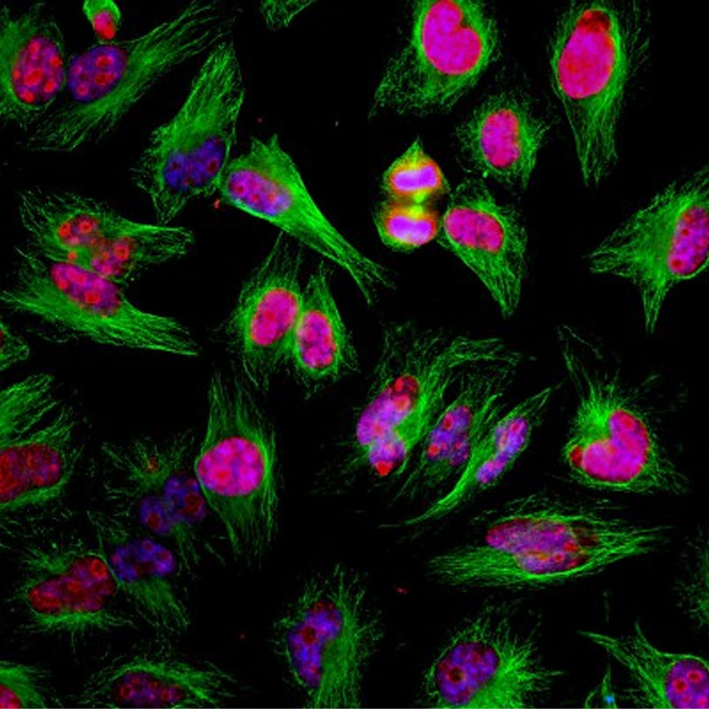
Immunofluorescent analysis of HeLa cells stained with chicken pAb to Ki67, CPCA-Ki67, dilution 1:2,000 in red, and mouse mAb to β-tubulin MCA-1B12, dilution 1:5,000 in green. The blue is DAPI staining of nuclear DNA. The CPCA-Ki67 antibody detects Ki67 protein predominantly expressed in nucleoli of cells in interphase and in mitotic cells Ki67 forms a coat around condensed chromosomes. Ki67 is not detected in cells in the quiescent G0 state. The MCA-1B12 antibody produces strong staining of cytoplasmic microtubules.
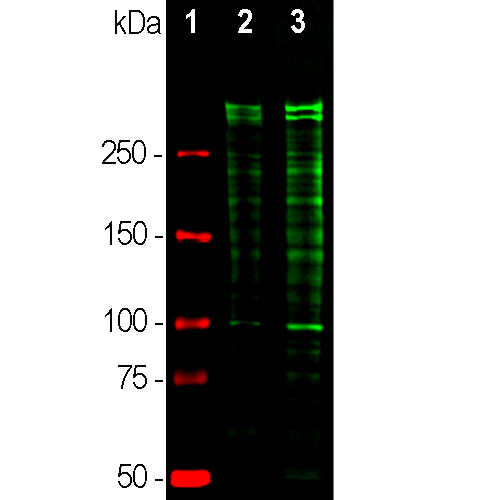
Western blot analysis of equal amounts of cell lysates probed with chicken pAb to Ki67, CPCA-Ki67, dilution 1:5,000, in green: [1] protein standard (red), [2] rapidly dividing HeLa cell cultures, [3] rapidly dividing HEK293 cell cultures. Strong double bands above 250kDa correspond to the two major Ki67 isoforms of molecular weight 345kDa and 395kDa. Since Ki67 is a rather unstable snd sjhort half life protein multiple proteolytic fragments of the two isoforms are also detected on the blot.
Chicken Polyclonal Antibody to Ki67, Ki-67
Cat# CPCA-Ki67
Price range: $120.00 through $800.00
The Ki67 protein was first discovered when researchers attempted to generate cancer cell specific monoclonal antibodies by injecting mice with nuclear preparations from Hodgkin’s lymphoma cells (1). They obtained a monoclonal antibody which recognized two large proteins of apparent molecular weight 345kDa and 395kDa. The clone was named Ki67 after Kiel, Germany where the original work was done and the number of the 96 well plate in which the clone was found. The two proteins were found to be heavily expressed in proliferating cells, but to be absent in quiescent cells, and later work showed that they were the product of a single gene. The presence of the Ki67 protein is frequently used as an indicator of cell proliferation and its level of expression is one of the most reliable biomarkers of proliferative status of cancer cells (2-5). Much research shows a correlation between Ki67 protein level and prognosis in cancer patients, when high Ki67 levels being associated with poorer outcomes (e.g. 6,7). The original Ki67 antibody and several others have become so widely used that a search for “(Ki67 or Ki-67) and antibody” in PubMed in August 2018 produced over 5,600 results. Recent studies show that Ki67 functions as a “biological surfactant”, which is essential for the fidelity of separation of condensed chromosomal DNA into the two daughter cells during cell division (8). This presumably explains the highly basic nature of Ki67, allowing a charge-based interaction with nucleic acids, the lack of this protein in non-dividing cells and the relative lack of protein sequence conservation.
The CPCA-Ki67 was made against a recombinant construct including amino acids 1,111-1,490 of the human sequence P46013, a region corresponding to 2nd, 3rd and 4th Ki67 type repeats. Although Ki67 is relatively poorly conserved in amino acid sequence, this antibody recognizes human, rat and mouse Ki67 on western blots. It can be used to visualize dividing cells and condensed chromosomes by IF, ICC and IHC. For IHC data select the “Additional Info” tab. iNote that the Ki67 proteins are very unstable and only expressed in large amounts in situations where many cells are dividing. As a result of the instability of the proteins there are usually numerous Ki67 fragments on western blots below the 395 and 345kDa. Mouse select image at left for larger view.

Chromogenic immunostaining of a formalin fixed paraffin embedded murine small intestine cross section with chicken pAb to Ki67, CPCA-Ki67, dilution 1:10,000, detected in DAB (brown) following the ABC method. Hematoxylin (blue) was used as the counterstain. Ki67 antibody specifically detects the nuclei of proliferating intestinal cells. Mouse select image at left for larger view.
1. Gerdes J, Schwab U, Lemke H, Stein H. Production of a mouse monoclonal antibody reactive with a human nuclear antigen associated with cell proliferation. Int. J. Cancer 31:13-20 (1983).
2. Kill IR, Faragher RGA, Lawrence K. Shall S. The expression of proliferation-dependent antigens during the lifespan of normal and progeroid human fibroblasts in culture. J. Cell Sci. 107:571-9 (1994).
3. Yerushalmi R, et al. Ki67 in breast cancer: Prognostic and predictive potential. Lancet Oncol. 11:174–83 (2010).
4. Josefsson A, et al. Low endoglin vascular density and Ki67 index in Gleason score 6 tumours may identify prostate cancer patients suitable for surveillance. Scand. J. Urol. Nephrol. 46:247–57 (2012).
5. Ishihara M, et al. Retrospective analysis of risk factors for central nervous system metastases in operable breast cancer: effects of biologic subtype and Ki67 overexpression on survival. Oncology. 84:135–140 (2013).
6. Cheang MC, et al. Ki67 Index, HER2 Status, and Prognosis of Patients With Luminal B Breast Cancer. J. Natl. Cancer Inst. 101:736-50 (2009).
7. Margulis V, et al. Multi-institutional validation of the predictive value of Ki-67 labeling index in patients with urinary bladder cancer. J. Natl. Cancer Inst. 101:114-9 (2009).
8. Cuylen S, et al. Ki-67 acts as a biological surfactant to disperse mitotic chromosomes .Nature. 535:308-12 (2016).
Related products


Recently Viewed Products
-
 Mouse Monoclonal Antibody to Neurofilament NF-L
Cat# MCA-1B11 Price range: $120.00 through $800.00 -
 Mouse Monoclonal Antibody to all Actin Isotypes
Cat# MCA-5J11 Price range: $120.00 through $800.00

