| Name: | Chicken Polyclonal Antibody to Ankyrin 3/Ankyrin G |
| Immunogen: | The C-terminal 398 amino acids of human ankyrin 3 expressed in and purified from E. coli |
| HGNC Name: | ANK3 |
| UniProt: | Q12955 |
| Molecular Weight: | 480, 270, and 190kDa |
| Host: | Chicken |
| Isotype: | IgY |
| Species Cross-Reactivity: | Human, rat, mouse, cow, monkey |
| RRID: | AB_2737591 |
| Format: | Purified antibody at 1mg/mL in 50% PBS, 50% glycerol plus 5mM NaN3 |
| Applications: | WB, IF/ICC, IHC |
| Recommended Dilutions: | WB: 1:5,000-1:10,000. IF/ICC 1:500-1:1,000. IHC not recommended |
| Storage: | Store at 4°C for short term, for longer term at -20°C. |
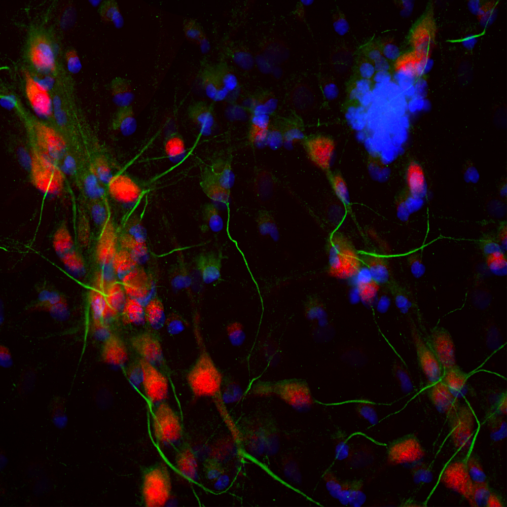
Immunofluorescent analysis of cortical neuron-glial cell culture from E20 rat stained with chicken pAb to ankyrin 3, CPCA-ANK3, dilution 1:2,000 in green, and costained with mouse mAb to FOX3/NeuN, MCA-1B7, dilution 1:2,000 in red. The blue is Hoechst staining of nuclear DNA. The CPCA-ANK3 antibody stains the axonal initial segments, while the FOX3/NeuN antibody reveals perikarya and nuclei of neurons.
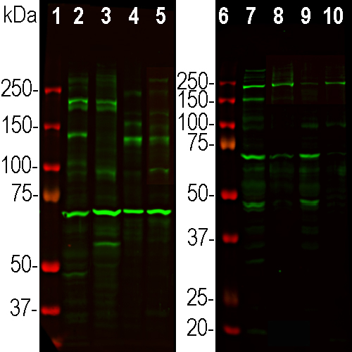
Western blot analysis of different tissue and cell lysates using chicken pAb to ankyrin 3, CPCA-ANK3, dilution 1:3,000, in green. [1, 6] Protein standard, [2] Rat brain, [3] Cow cortex, [4] HEK293 cells, [5] COS-1 cells, [7] Rat cortex, [8] rat cortex membrane enriched fraction, [9] mouse cortex, [10] mouse cortex membrane enriched fraction. Bands at 60-190kDa represent ankyrin 3 splice variants and proteolytic fragments. Larger ankyrin 3 isoforms of 270kDa and 480kDa can be seen on longer exposure of the blots.
Chicken Polyclonal Antibody to Ankyrin 3/Ankyrin G
Cat# CPCA-ANK3
Price range: $150.00 through $1,000.00
The first ankyrin protein was isolated from red blood cell membranes and was found to be responsible for anchoring the spectrin cytoskeleton to the plasma membrane. Subsequently this protein was named ankyrin 1 and a close homolog was named ankyrin 2, though the two proteins are also known as ankyrin R and ankyrin B, for erythrocyte and brain respectively (2). Both proteins are large at about 200kDa. A third member of the protein family was discovered and called ankyrin 3, also known as ankyrin G, some forms of which are much larger in molecular size, up to 480kDa. G refers to giant or general, as this protein is large and widely expressed (2). All three ankyrins have an N-terminal segment composed of 22 tandem repeats each of 33 amino acids which have been named ankyrin type repeats. A subset of these repeats are responsible for binding the ankyrin proteins to various membrane proteins, and repeats of this kind are found in many other proteins and generally mediate specific protein-protein interactions (3). The middle region of the molecules contain the spectrin binding activity and the C-terminal contains a DEATH domain and some other sequence which is variable between the three ankyrins. DEATH domains are involved in activating apoptotic pathways, and are found in many molecules of known apoptotic function such as the TNF receptor and Fas/Apo1 (4). The much larger size of ankyrin 3 is due to a sequence which may be inserted within this C-terminal region. The ankyrin 3 gene may produce a protein of 480kDa while other transcripts produce 270kDa and 190kDa proteins. Defects in the ankyrin gene are associated with various human disorders (5,6). Ankyrin 3 is expressed in the axon initial segment and the nodes of Ranvier in the nervous system so appropriate antibodies are useful to identify these regions (7). The function of ankyrin 3 appears to be to specifically localize channels and other cytoskeletal proteins at these regions.
The CPCA-ANK3 antibody was made against the C-terminal 398 amino acid of human isotype 1 in NP_066267.2. This segment is expressed by all ankyrin 3 three isotypes and contains the DEATH domain sequence. The antibody works well for IF and ICC, but is not recommended for IHC. EnCor also supplies a mouse monoclonal and a rabbit polyclonal to this protein made using the same immunogen, MCA-2A8 and RPCA-ANK3.
The first ankyrin protein was isolated from red blood cell membranes and was found to be responsible for anchoring the spectrin cytoskeleton to the plasma membrane. Subsequently this protein was named ankyrin 1 and a close homolog was named ankyrin 2, though the two proteins are also known as ankyrin R and ankyrin B, for erythrocyte and brain respectively (2). Both proteins are large at about 200kDa. A third member of the protein family was discovered and called ankyrin 3, also known as ankyrin G, some forms of which are much larger in molecular size, up to 480kDa. G refers to giant or general, as this protein is large and widely expressed (2). All three ankyrins have an N-terminal segment composed of 22 tandem repeats each of 33 amino acids which have been named ankyrin type repeats. A subset of these repeats are responsible for binding the ankyrin proteins to various membrane https://files.encorbio.com/product/cpca-ank3/proteins, and repeats of this kind are found in many other proteins and generally mediate specific protein-protein interactions (3). The middle region of the molecules contain the spectrin binding activity and the C-terminal contains a DEATH domain and some other sequence which is variable between the three ankyrins. DEATH domains are involved in activating apoptotic pathways, and are found in many molecules of known apoptotic function such as the TNF receptor and Fas/Apo1 (4). The much larger size of ankyrin 3 is due to a sequence which may be inserted within this C-terminal region. The ankyrin 3 gene may produce a protein of 480kDa while other transcripts produce 270kDa and 190kDa proteins. Defects in the ankyrin gene are associated with various human disorders (5,6). Ankyrin 3 is expressed in the axon initial segment and the nodes of Ranvier in the nervous system so appropriate antibodies are useful to identify these regions (7). The function of ankyrin 3 appears to be to specifically localize channels and other cytoskeletal proteins at these regions.
The CPCA-ANK3 antibody was made against the C-terminal 398 amino acid of human isotype 1 in NP_066267.2. This segment is expressed by all ankyrin 3 three isotypes and contains the DEATH domain sequence. EnCor also supplies a mouse monoclonal and a rabbit polyclonal to this protein, MCA-2A8 and RPCA-ANK3. Mouse select image above left for larger view.
1. Bennett, V. Adaptors between divese plasma membrane proteins and the cytoplasm. J. Biol. Chem. 287:8703-6 (1992).
2. Kordeli, E, Lambert, S, Bennett, V. AnkyrinG. J. Biol. Chem. 270:2352-9 (1995).
3. Mosavi LV, Cammett TJ, Desrosiers DC, Peng Z. The ankyrin repeat as molecular architecture for protein recognition. Protein Sci. 13:1435-48 (2004).
4. Feinstein E, et al. The death domain: a module shared by proteins with diverse cellular functions. Trends Biochem. Sci. 20:342-4 (1995).
5. Cunha SR, Mohler PJ. Ankyrin protein networks in membrane formation and stabilization. J. Cell Mol. Med. 13:4364-76 (2009).
6. Lopez AY, et al. Ankyrin-G isoform imbalance and interneuronopathy link epilepsy and bipolar disorder. Mol. Psychiatry 22:1464-72 (2017).
7. Alshammari MA, Alshammari TK, Laezza F. Improved Methods for Fluorescence Microscopy Detection of Macromolecules at the Axon Initial Segment. Front. Cell Neurosci. 2016 10:5 (2016).


